
50-year-old man with history of elevated d-dimer with recent history of chest discomfort presents for evaluation for pulmonary embolism
No pulmonary embolism was identified
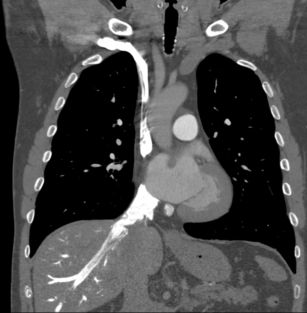
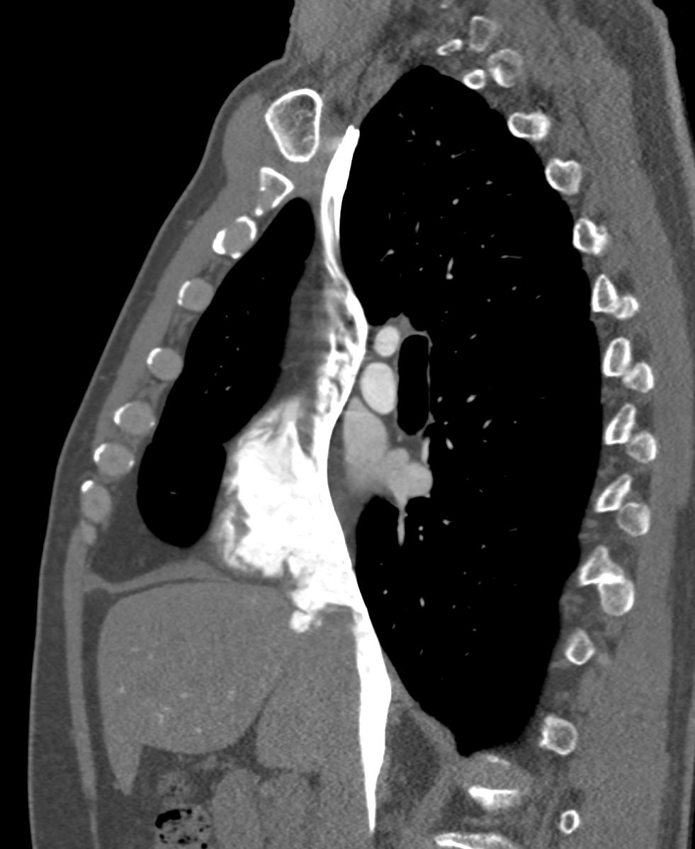
There was however significant stasis of systemic venous inflow into the right atrium and right ventricle characterized by almost complete stasis of the contrast column in the IVC and extension of the contrast into the smallest hepatic veins in the dependent portion of the liver posteriorly. There was also reflux of contrast down the azygos vein
The right atrium and right ventricle were normal in size. The left atrium and the left ventricle were slightly enlarged with bulging of the inter-atrial septum toward the right atrium suggesting that the left atrial pressure was elevated.
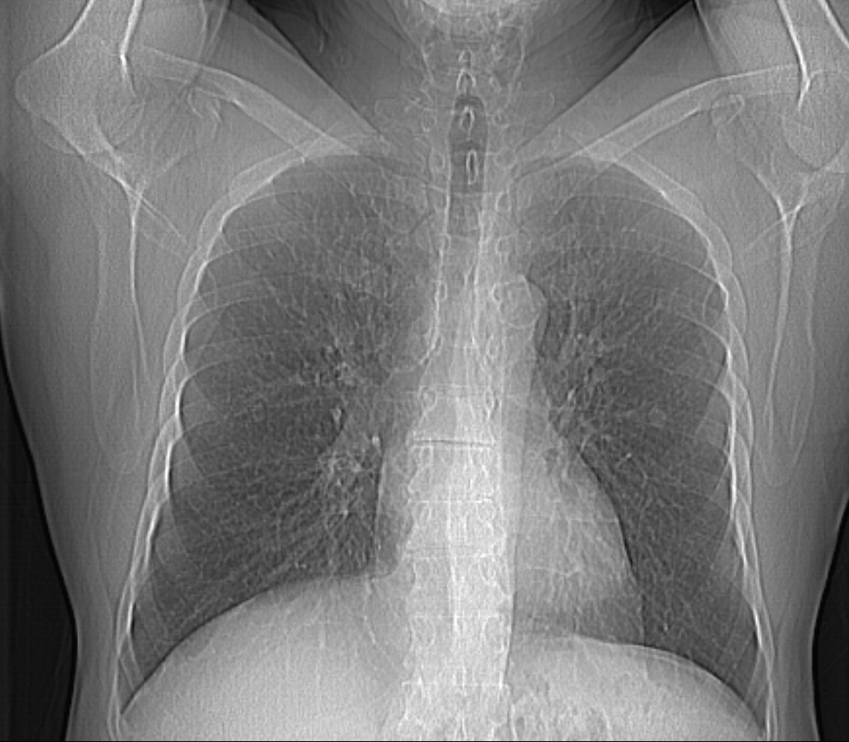
Scout film of the chest prior to the CT scan is normal
Ashley Davidoff MD TheCommonVein.net
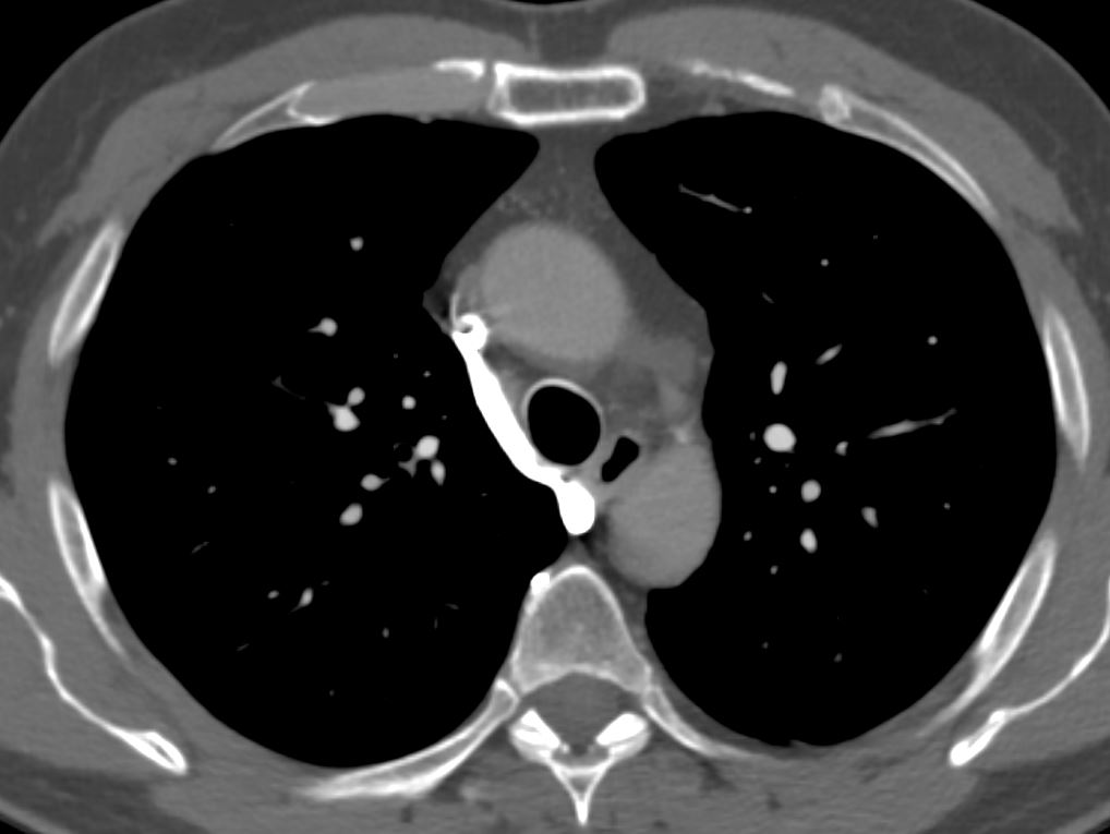
CT scan shows reflux into the azygous vein
Ashley Davidoff MD TheCommonVein.net
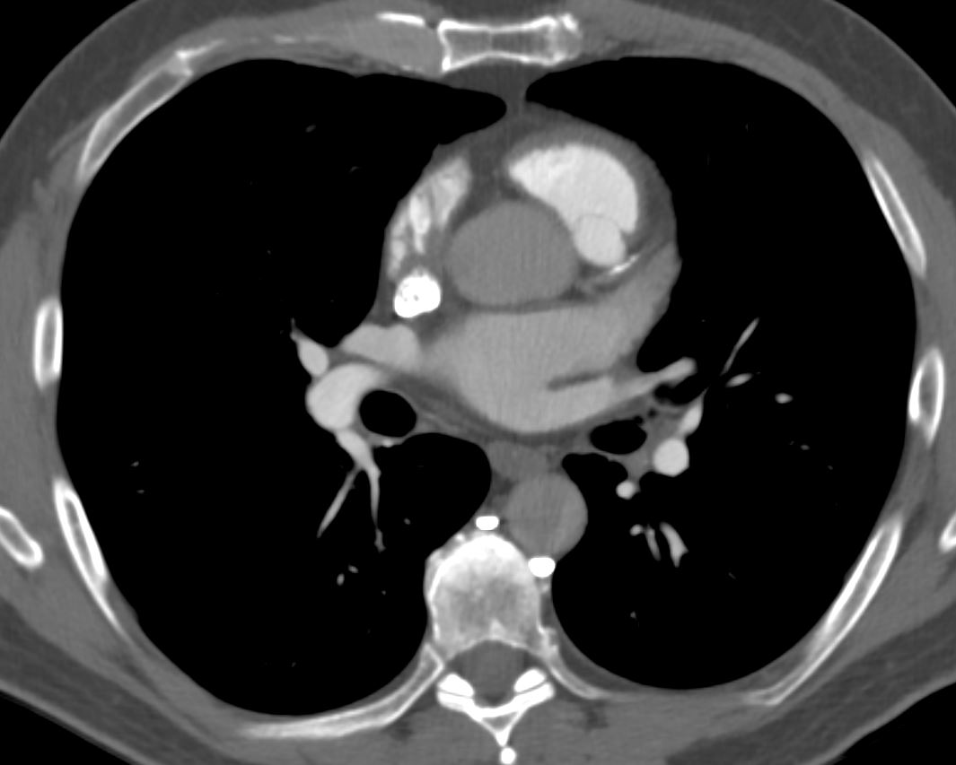
CT scan shows mild LAD calcification
Ashley Davidoff MD TheCommonVein.net
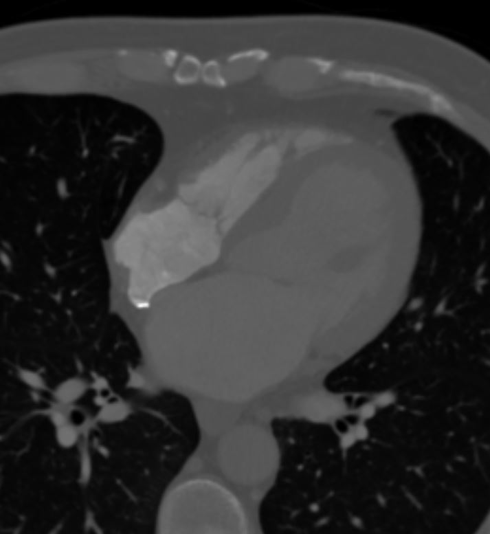
CT scan normal to small right sided chambers, enlarged left atrium and left ventricle, bulging of the septum primum to the right indicating that left atrial pressures are higher than the right
Ashley Davidoff MD TheCommonVein.net
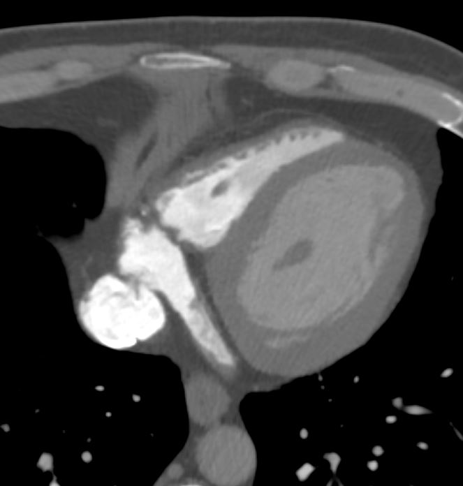
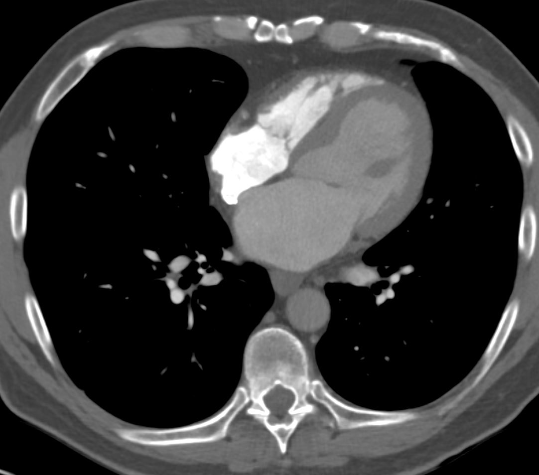
CT scan small right ventricle, dilated left ventricle, enlarged IVC and dilated coronary sinus.
Ashley Davidoff MD TheCommonVein.net
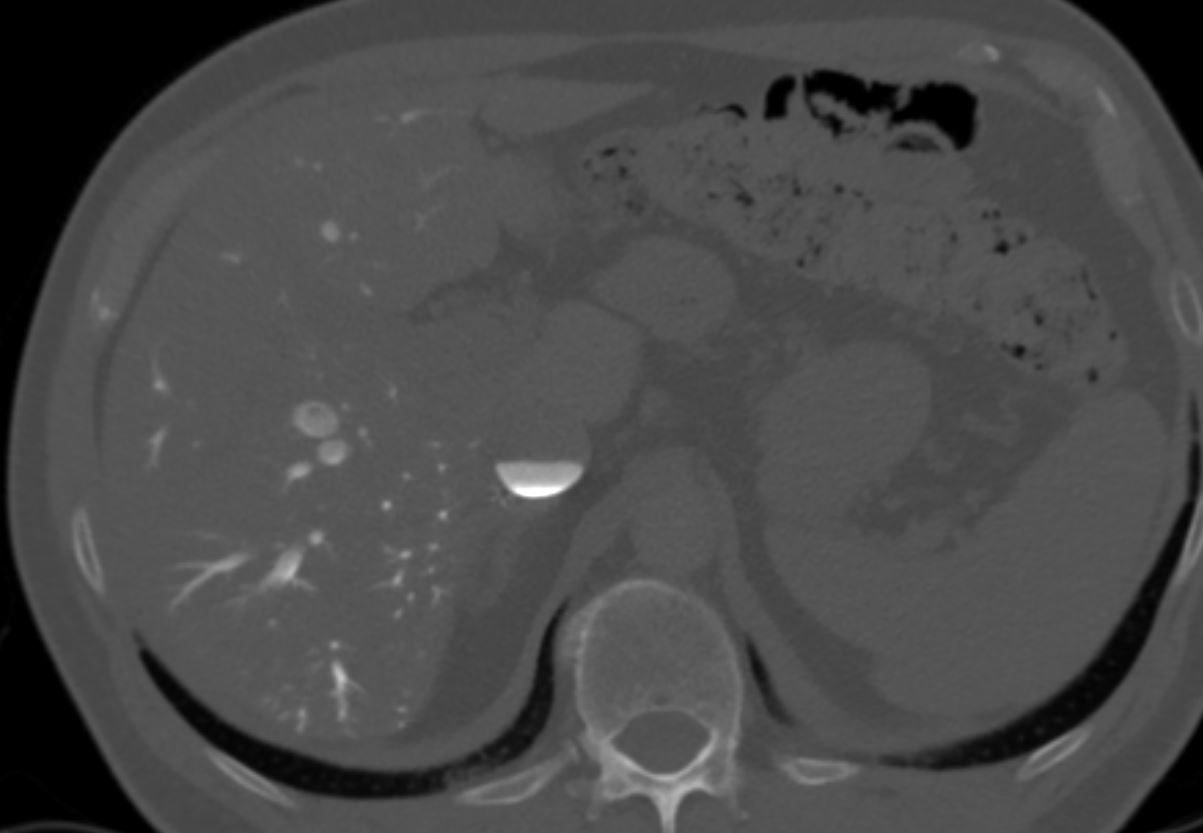
There is significant stasis of systemic venous inflow In this CT image there is almost complete stasis of the contrast column in the IVC with a contrast blood level that characterizes the “IVC level” sign There is extension of the contrast into the smallest
hepatic veins in the dependent portion of the liver posteriorly.
Ashley Davidoff MD TheCommonVein.net
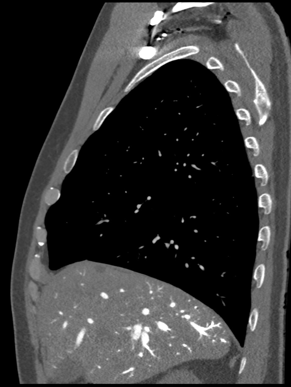
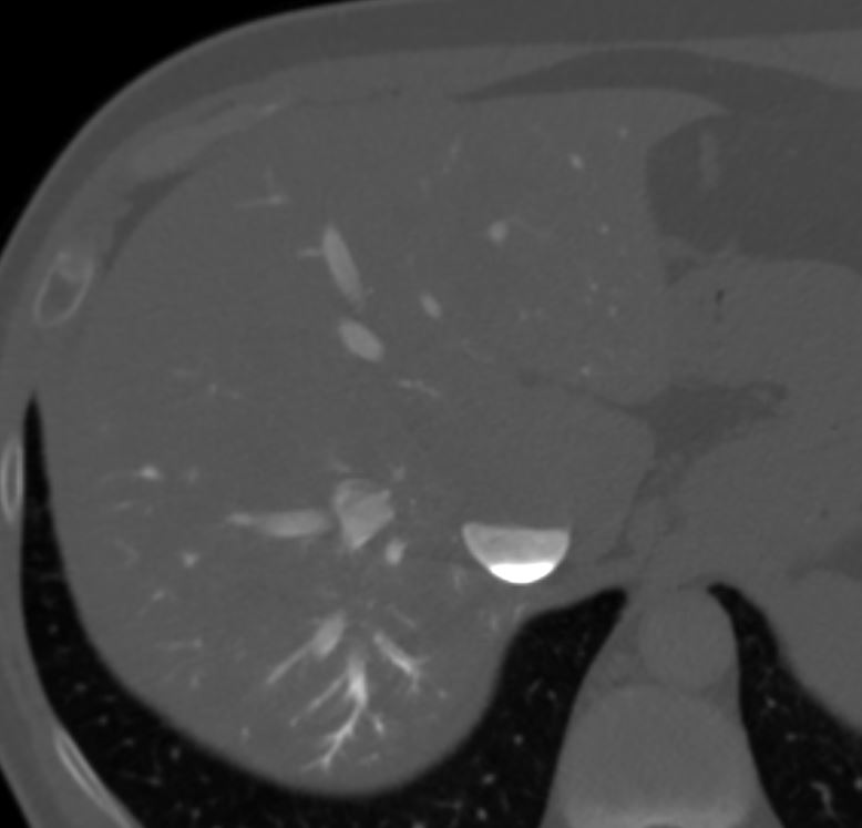
There is significant stasis of systemic venous inflow In this CT image there is extension of the contrast into the smallest hepatic veins in the dependent portion of the liver posteriorly.
Ashley Davidoff MD TheCommonVein.net
There is significant stasis of systemic venous inflow In this CT image there is almost complete stasis of the contrast column extends into the smallest
hepatic veins in the dependent portion of the liver posteriorly indicating significant stasis.
Ashley Davidoff MD TheCommonVein.net
There is significant stasis of systemic venous inflow In this CT image there is almost complete stasis of the contrast column extends into the smallest
hepatic veins in the dependent portion of the liver posteriorly indicating significant stasis.
Ashley Davidoff MD TheCommonVein.net
Venous findings suggest limitation of flow into the right atrium, that may be seen in the setting of shock or significant pericardial constriction or cardiac tamponade. However there was no report of acute hemodynamic compromise while the patient was in the scanner nor with subsequent communication with nurse who had communicated with the patient. Theoretically, severe Valsalva maneuver could. also limit flow into the right atrium could theoretically also result in a similar appearance but it would be very unusual