
- Normally,
- vessels in the lung bases are
- larger and
- more numerous than
- lung apices.
- Due to
- secondary to the effects of gravity and
- anatomically larger volume of the lungs at the base.
- vessels in the lung bases are
- The pulmonary vascular bed is a
- low resistance,
- low pressure system
- unique
- the only circulation that can
- receive the entire ventricular output with
- one systolic contraction.
- cardiac output can vary from a
- normal of approximately
- 5 liters perminute1 to a
- 40 liters per minute during strenuous exercise.
- accommodating these large increases
- with little change in pressure.
- ie lungs have significant reserve capacity
- with an ability to
- recruit more circulation when needed
- by opening previously nonperfused
vascular channels,
- normal of approximately
- the only circulation that can

Ashley Davidoff MD
TheCommonVein.net
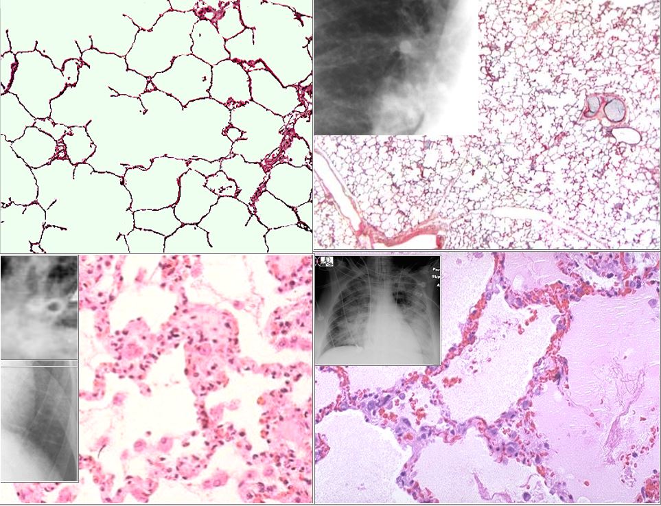
The top left image is the a histological section of normal alveoli and normal wall and interstitium. Heart failure occurs when the left ventricular end diastolic pressure rises. There are 3 basic phases of heart failure. in the first phase (top right) the LVEDP rises above 12 mmHg and on an upright CXR there is equalization of the size of the vessels going to the upper lobes and lower lobes. As the LVEDP goes above about 15-18 mm Hg there is cephalization of the vessels and the upper lobe vessels are larger than the lower lobe vessels.
The second phase of interstitial edema (bottom left) occurs when the intravascular hydrostatic pressure exceeds the intravascular oncotic pressure and this occurs when the LVEDP goes above 25 mm Hg. Fluid accumulates in the alveolar walls and interstitium and the wall becomes thicker with fluid, and the lymphatics and interlobular septa are distended.
The last phase of alveolar edema (lower right) occurs when the pressure exceeds 35 mmHg and the fluid leaks into the alveoli .
Ashley Davidoff MD
- In supine position
-
- upper and lower lung fields show equalization
-
There is a tremendous amount of reserve in the vascular flow of the lungs and they
The earliest recognizable level of redistribution occurs when perfusion of upper and lower lung zones is equal.’
- As redistribution becomes more pronounced, the
- size and
- number of vessels in upper lung zones
- exceed that seen in lower lung zones.
- vasoconstriction may occur in the lower lung zone
- Note
- lateral view can be extremely helpful to identify
- the enlargement of the pulmonary vessels
- Limitations
- Suboptimal inspiration
- may result in apparent
- redistribution of pulmonary blood flow
Links and References
Kramer B et al Hemodynamic Differences Between Supine and
Upright Exercise in Patients with Congestive Heart Failure
Ravin, Carl Radiographic analysis of pulmonary vascular redistribution: A Review