
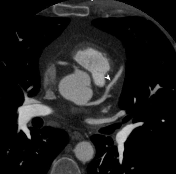
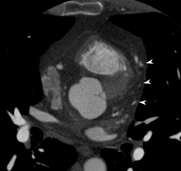
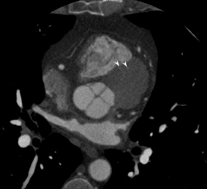
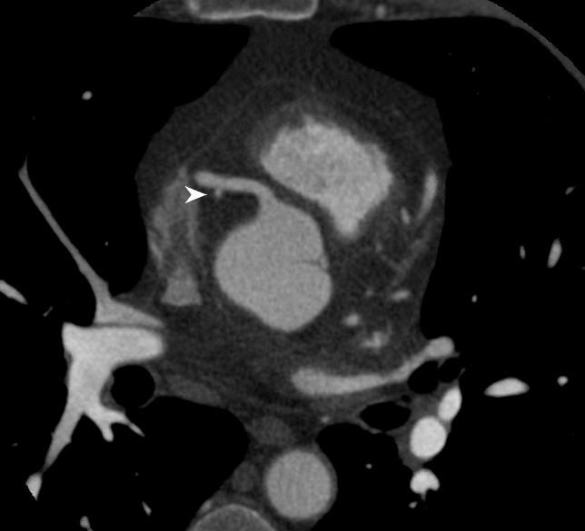
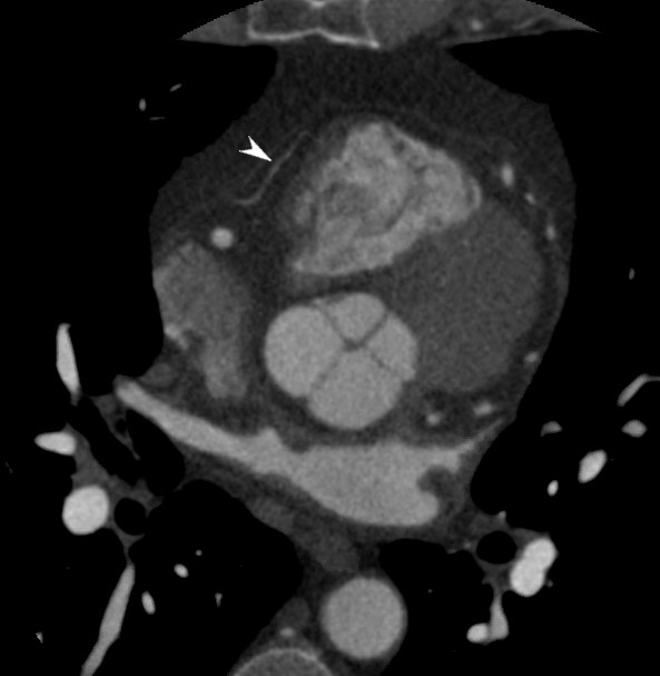
Left Main
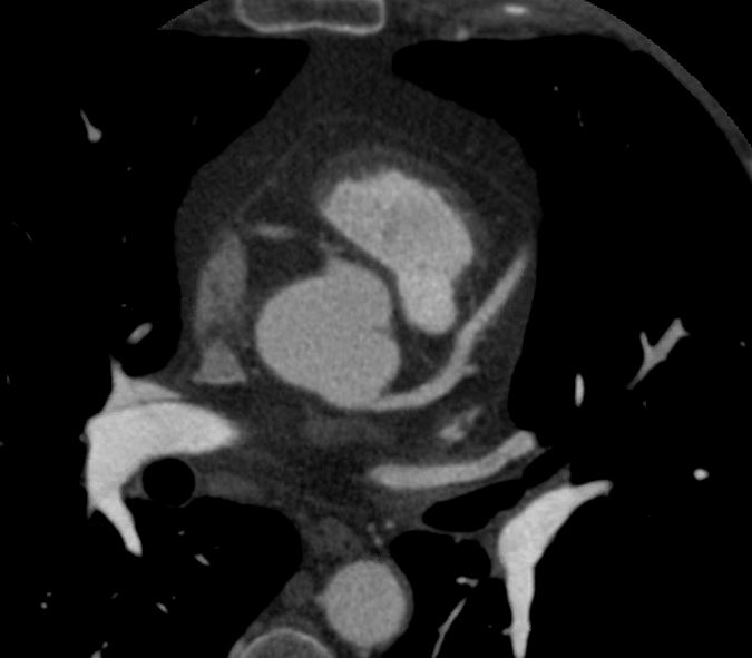
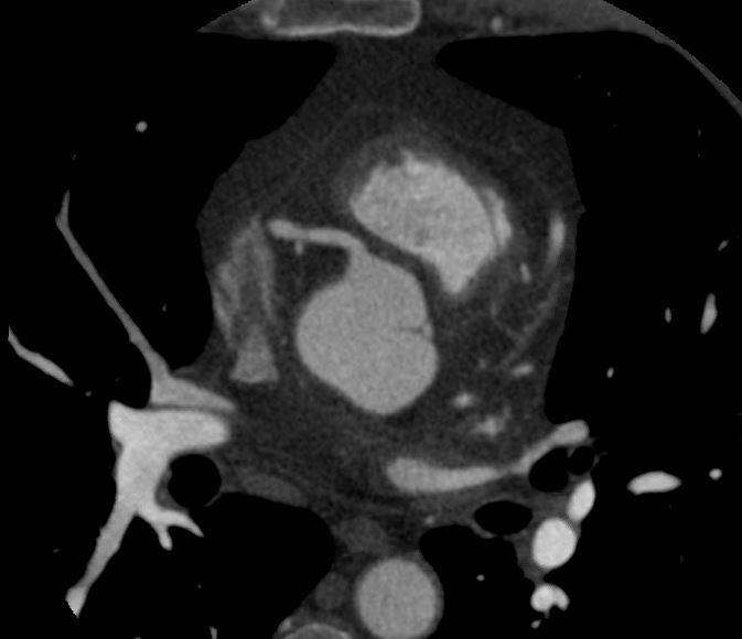
Normal axial CT of the left main coronary artery (LM) showing its origin from the left coronary cusp which is posterior but slightly superior to the right coronary cusp
Ashley Davidoff MD
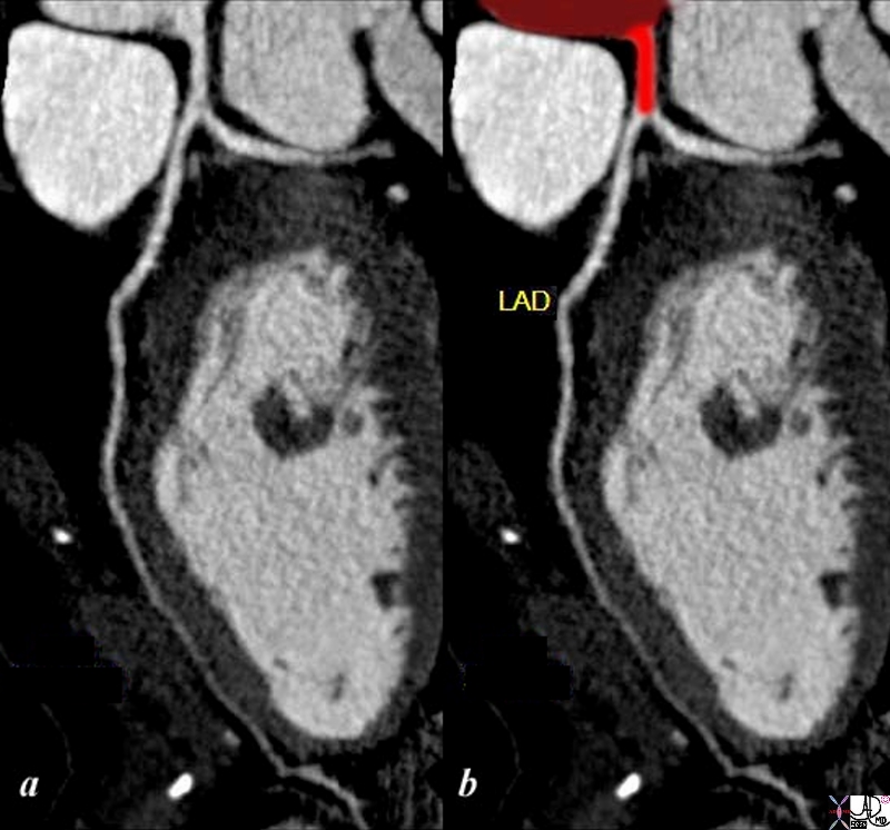
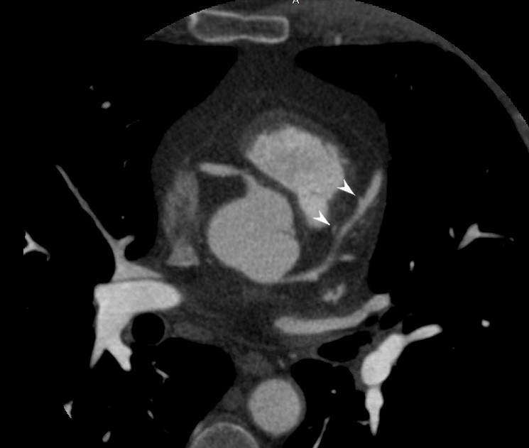
Reconstruction of the CTA of the right left coronary artery shows LM (overlaid in red) and the LAD. They are both normal
Ashley Davidoff MD thecommonvein.net
87261c02.8s
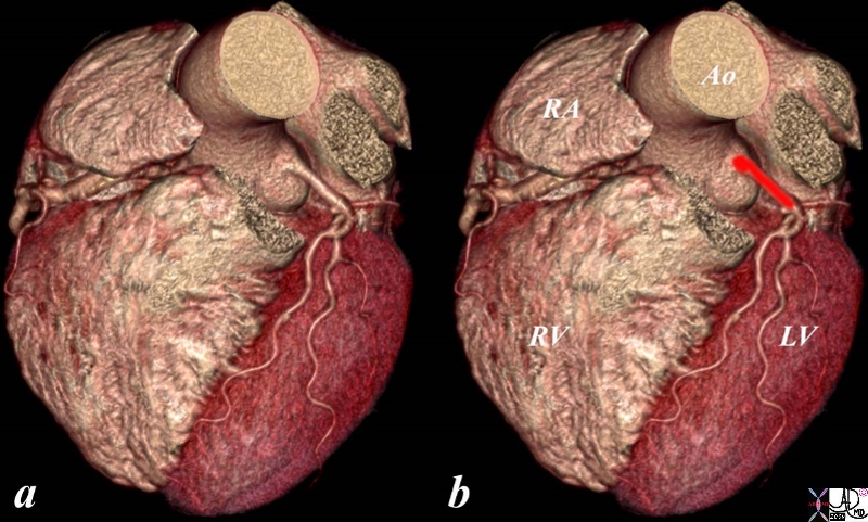
Reconstruction of the CTA of the right left coronary artery shows LM (overlaid in red) and the LAD. They are both normal
Ashley Davidoff MD thecommonvein.net
87255c02.8s
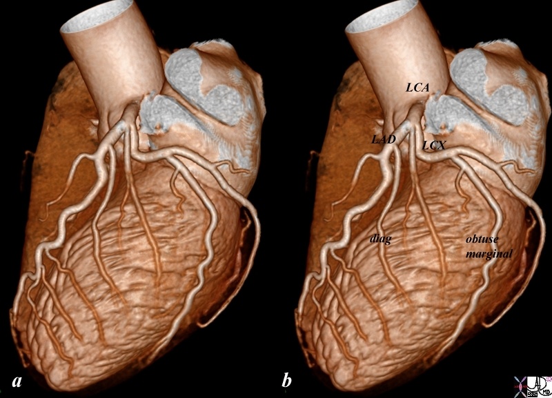
Reconstruction of the CTA of the left left coronary artery shows normal LM , LAD and diagonals and circumflex and obtuse marginals .
Ashley Davidoff MD thecommonvein.net
87260b01.8s
Normal axial CT of the left anterior descending coronary artery
Ashley Davidoff MD thecommonvein.net
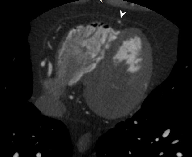
Normal axial CT of the left anterior descending coronary artery
Ashley Davidoff MD thecommonvein.net
Reconstruction of the CTA of the right coronary artery in the AP projection showing the acute marginal branch. Note the LAD and some diagonal vessels shown as well
Ashley Davidoff MD thecommonvein.net
45167
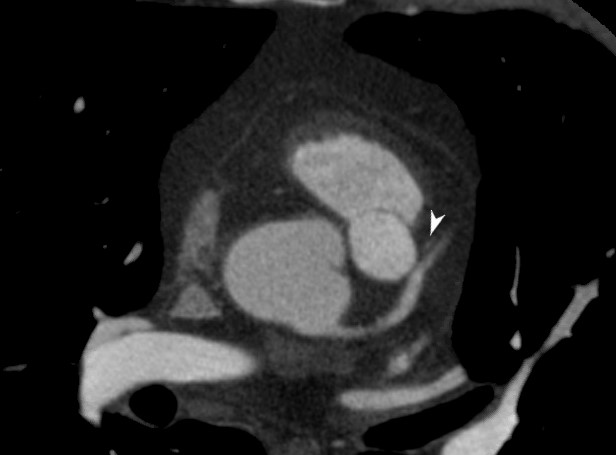
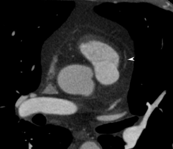
Diagonals
Normal axial CT of the left anterior descending coronary artery showing the diagonal arteries supplying the anterior wall of the left ventricle (LV)
Ashley Davidoff MD thecommonvein.net
Normal axial CT of the left anterior descending coronary artery showing the diagonal arteries supplying the anterior wall of the left ventricle (LV)
Ashley Davidoff MD thecommonvein.net
Normal axial CT of the left anterior descending coronary artery showing the diagonal arteries supplying the anterior wall of the left ventricle (LV)
Ashley Davidoff MD thecommonvein.net
Reconstruction of the CTA of the left anterior descending coronary artery showing the diagonal anterolateral wall of the left ventricle (LV)
Ashley Davidoff MD thecommonvein.net
45168
Septals
Normal axial CT of the septal coronary arteries showing their origin from the LAD. They quickly enter the myocardium and are small and are therefore usually impossible to visualise
Ashley Davidoff MD thecommonvein.net

Normal axial CT of the septal coronary arteries showing their origin from the LAD. They quickly enter the myocardium and are small and are therefore usually impossible to visualise
Ashley Davidoff MD thecommonvein.net

Normal axial CT of the septal coronary arteries showing their origin from the LAD. They quickly enter the myocardium and are small and are therefore usually impossible to visualise
Ashley Davidoff MD thecommonvein.net
Normal axial CT of the septal coronary arteries showing their origin from the LAD. They quickly enter the myocardium and are small and are therefore usually impossible to visualise
Ashley Davidoff MD thecommonvein.net
Conal Artery
Ashley Davidoff MD thecommonvein.net
Ashley Davidoff MD thecommonvein.net
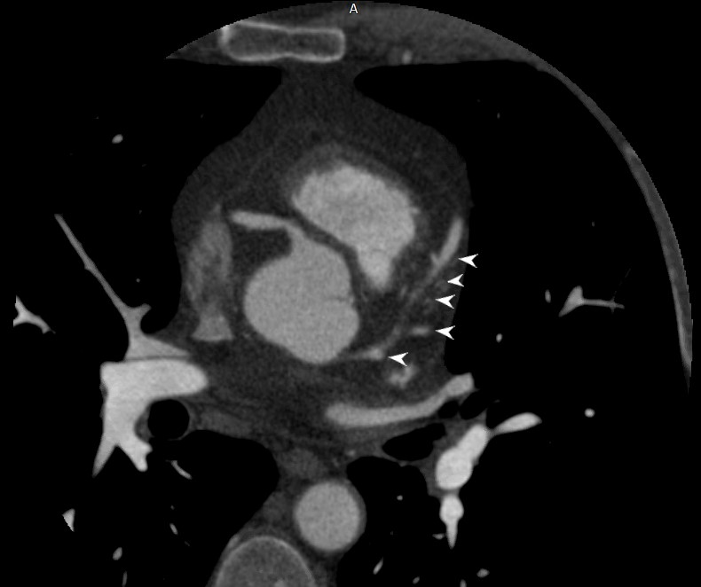
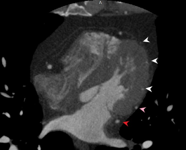
Circumflex Coronary Artery and Obtuse Marginals
Reconstruction of the CTA of the circumflex coronary artery showing the obtuse marginals supplying the posterolateral wall of the left ventricle (LV)
Ashley Davidoff MD thecommonvein.net
45168b01.8s
Reconstruction of the CTA of the distal circumflex and distal right coronary arteries in the posterior aspect of the heart showing the obtuse marginals supplying the posterior wall of the left ventricle (LV) and the distal RCA supplying the PDA and crossing over to give rise to the posterior left ventricular branch
Ashley Davidoff MD thecommonvein.net
45170
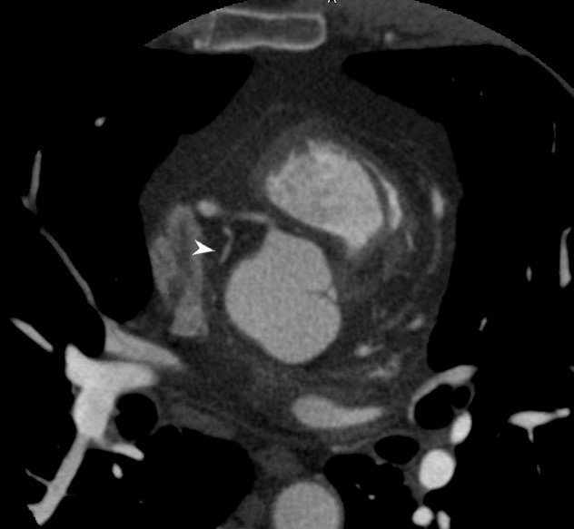
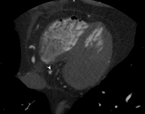
Right Coronary Artery
Normal axial CT of the right coronary artery showing its origin from the right coronary cusp which is anterior and slightly inferior to the left coronary cusp
Ashley Davidoff MD
Normal axial CT of the right coronary artery showing its origin from the right coronary cusp which is anterior and slightly inferior to the left coronary cusp
Ashley Davidoff MD
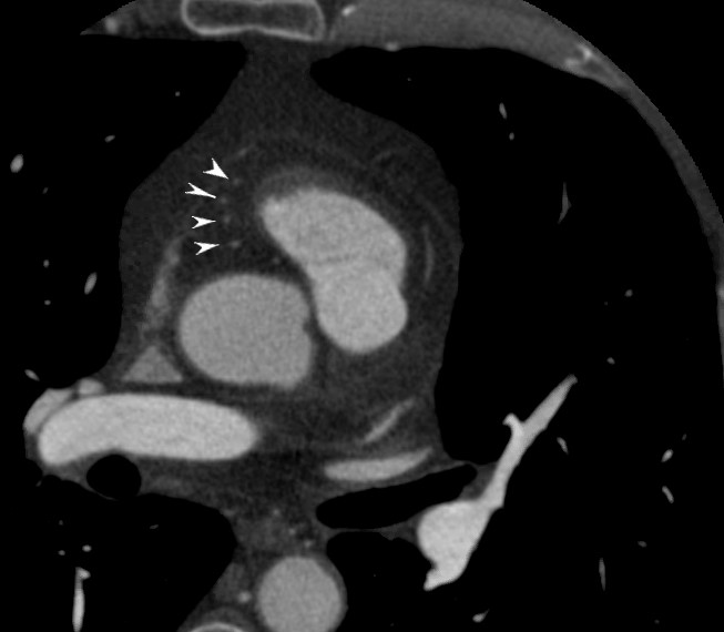
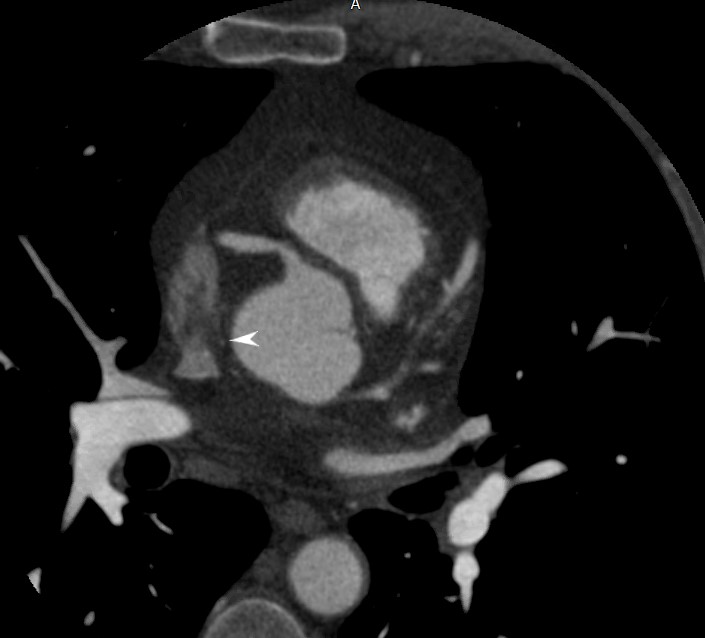
Conal Artery off the RCA – Arc of Vieussens
Normal axial CT of the right coronary artery showing the conal artery supplying the conus of the right ventricular outflow tract (RVOT)
Ashley Davidoff MD
Ashley Davidoff MD
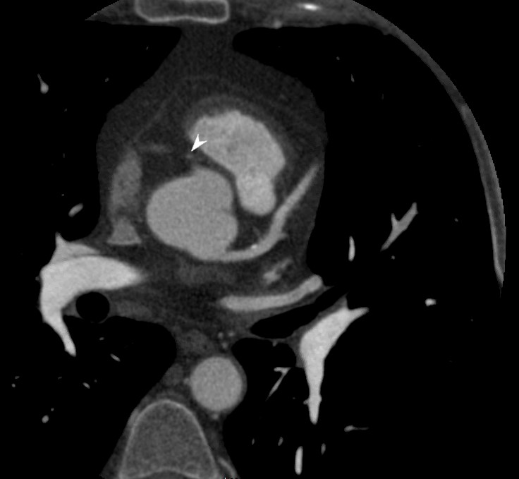
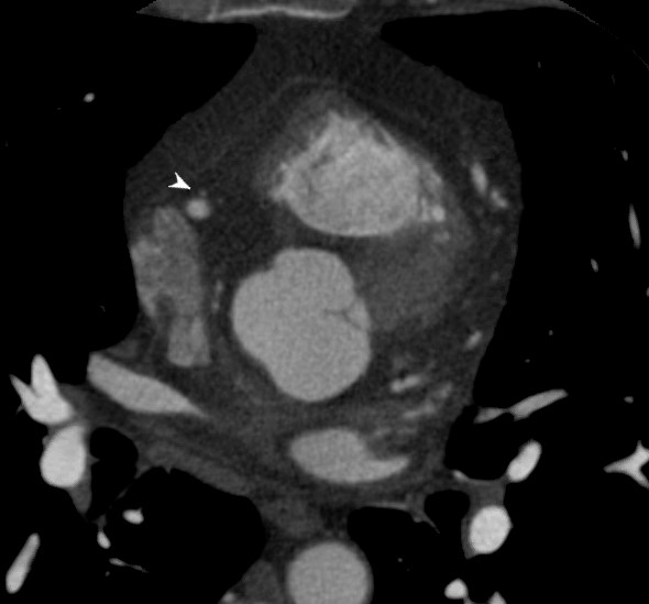
SA Nodal Artery off the proximal RCA
Normal axial CT of the right coronary artery showing the SA – Nodal artery supplying the SA Node at the SVC RA junction
Ashley Davidoff MD
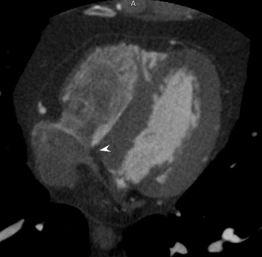
Acute Marginal Artery off the proximal RCA
Acute Marginal Artery off the proximal RCA
Normal axial CT of the right coronary artery showing the acute marginal artery of the right ventricle (RV)
Reconstruction of the CTA of the right coronary artery in the AP projection showing the acute marginal branch. Note the LAD and some diagonal vessels shown as well
Ashley Davidoff MD thecommonvein.net
45167
Ashley Davidoff MD
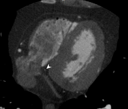
AV nodal Artery off the Distal RCA at the Crux of the Heart
Normal axial CT of the right coronary artery showing the AV nodal artery off the distal RCA at the crux of the heart
Ashley Davidoff MD

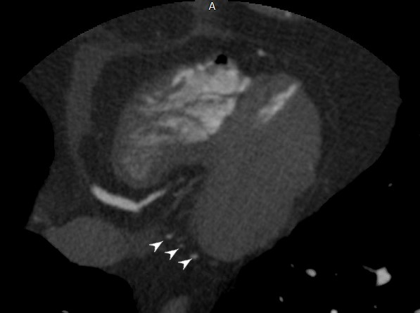
Posterior Left Ventricular Branches off the Distal RCA Supplying the Posterior Aspect of the LV
Normal axial CT of the right coronary artery showing the posterior left ventricular branches off the distal RCA supplying the posterior aspect of the LV
Ashley Davidoff MD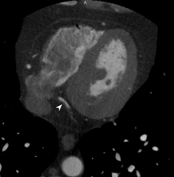
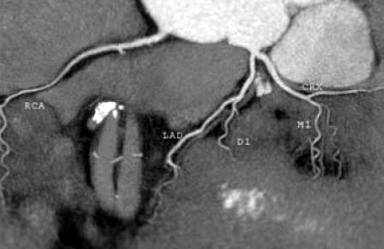
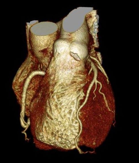
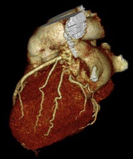
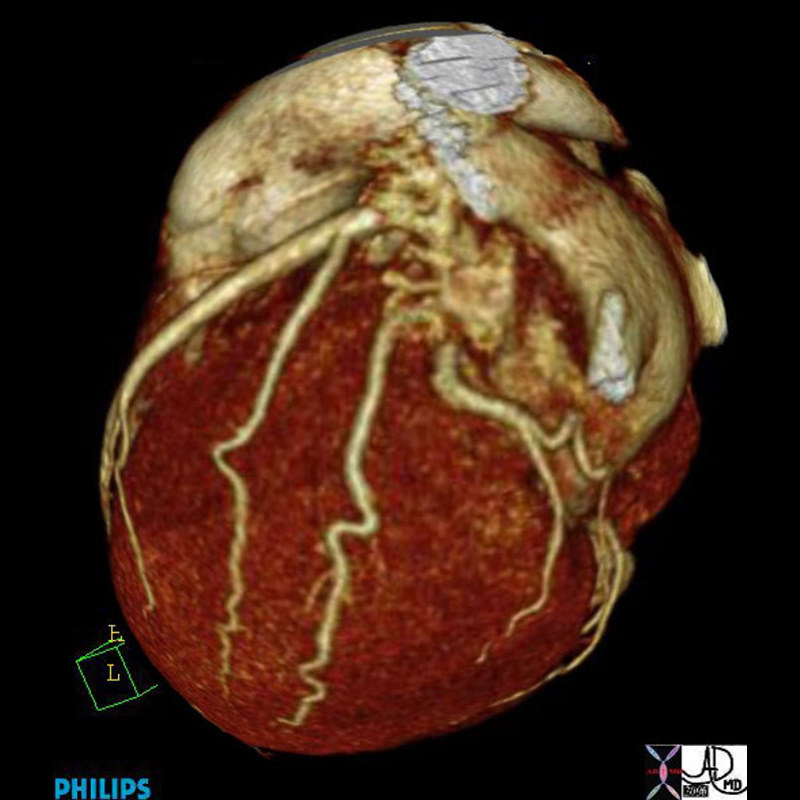
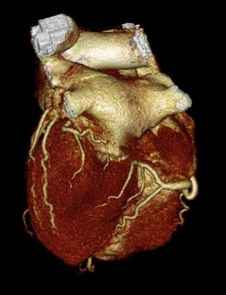
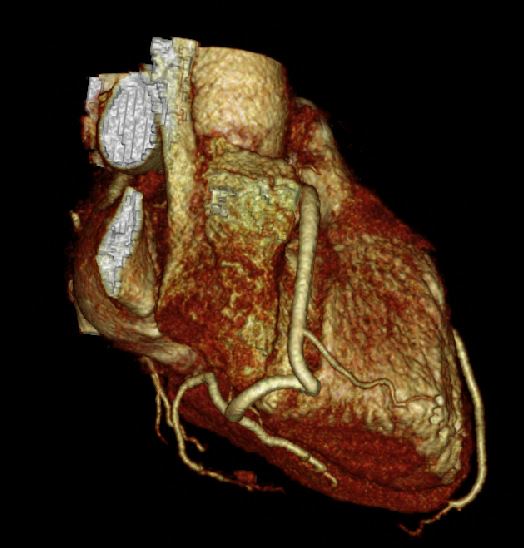
Reconstruction of the coronary arteries shows normal left main LAD, circumflex and RCA
Ashley Davidoff thecommonvein.net
44131.5
-
Links and References
-
TCV
LCA-