
- Motion is the most common caused by
- patient,
- cardiac, or
- respiratory motion.
- Attempt at solutions
- Cardiac motion artifacts
-
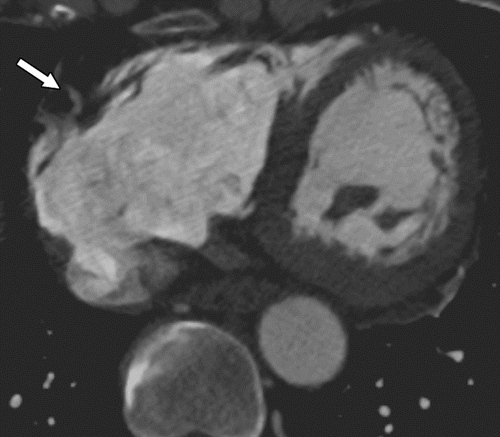
Cardiac motion artifacts. (a) Axial CT image shows blurring and winged appearance of the right coronary artery (arrow).
Kalisz K et al Artifacts at Cardiac CT: Physics and Solutions RadioGraphicsVol. 36, No. 7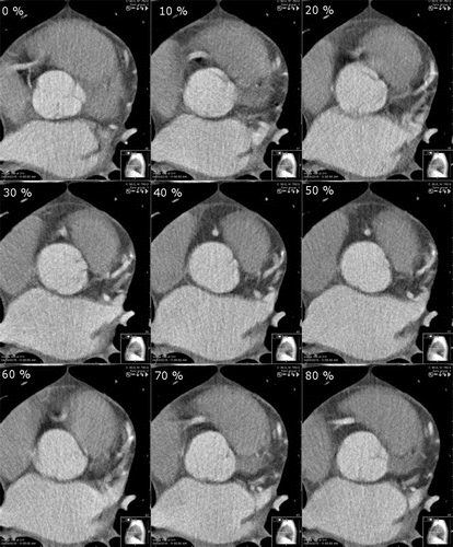
Axial CT images obtained at different phases of the cardiac cycle (from 0% to 80% R-R interval) show that the right coronary artery has varying degrees of motion in each of the cardiac phases. In this patient, the motion is least in the 50% and 80% phases of the R-R interval.
Kalisz K et al Artifacts at Cardiac CT: Physics and Solutions RadioGraphicsVol. 36, No. 7 - can be reduced by
- decreasing the heart rate and variability and the
- duration of data acquisition;
- adjusting the placement of the data window within a cardiac cycle; (diastole
- performing single-heartbeat scanning; and
- using multisegment reconstruction,
- motion-correction algorithms, and
- electrocardiographic editing.
- Respiratory motion artifacts can be minimized with
- proper breath holding and
- shortened scan duration.
- Partial volume averaging is
- caused by the
- averaging of attenuation values from all tissue contained within a voxel and
- reduced by
- improving the spatial resolution,
- using a higher x-ray energy, or
- displaying images with a wider window width.
- caused by the
- Beam-hardening artifacts are
-
- caused by the
- polyenergetic nature of the x-ray beam and can be
- reduced by using
- x-ray filtration,
- applying higher-energy x-rays,
- altering patient position,
- modifying contrast material protocols, and
- applying certain reconstruction algorithms.
- caused by the
- Metal artifacts are complex and
- have multiple causes,
- including x-ray scatter,
- underpenetration, motion, and
- attenuation values that exceed the typical dynamic range of Hounsfield units.
- have multiple causes,
- Quantum mottle or noise is
- caused by
- insufficient penetration of tissue and can be
- improved by
- increasing the tube current or peak tube potential,
- reconstructing thicker sections,
- increasing the rotation time,
- using appropriate patient positioning, and
- applying iterative reconstruction algorithms.
- caused by
-
Links and References
Kalisz K et al Artifacts at Cardiac CT: Physics and Solutions RadioGraphicsVol. 36, No. 7