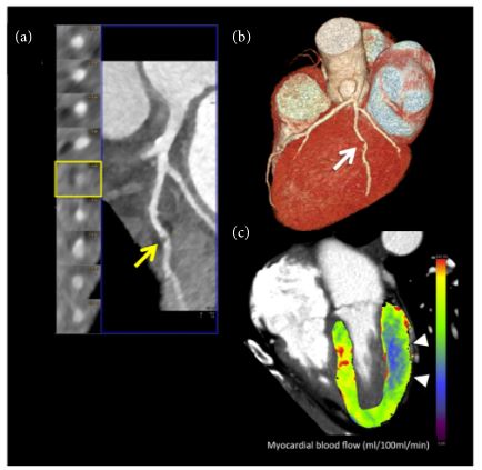
Dynamic CTP imaging. 67-year-old obese female patient with history of hyperlipidemia and smoking with suspected coronary artery disease. (a) Curved multiplanar reformation of coronary CT angiography data showed eccentric noncalcified plaque of the main obtuse marginal branch (OM) causing focal critical stenosis (>70% luminal narrowing), arrow. (b) Three-dimensional volume-rendering reconstruction confirmed the severe coronary artery stenosis of the OM (arrow). (c) Three-dimensional color-coded 4-chamber CT perfusion map image derived from the time-resolved dynamic acquisition during stress with the shuttle mode shows extensive perfusion defects in the territory of the OM (basal-middle lateral wall), color-coded in blue, arrowheads. The colors of the myocardium are coded according to the flow values with red, green, and yellow representing higher flow values than blue. The corresponding value of the hemodynamic parameters derived from the time-attenuation curves (TACs) demonstrates a significant reduction of myocardial blood flow in the territory of the OM, consistent with inducible ischemia. Absolute myocardial blood flow was 61.6 mL/100 mL/min and 118.2 mL/100 mL/min in the OM and remote myocardium (septal wall) territories, respectively. Seitun S, et al CT Myocardial Perfusion Imaging: A New Frontier in Cardiac Imaging BioMed Research International / 2018 Article ID 7295460