
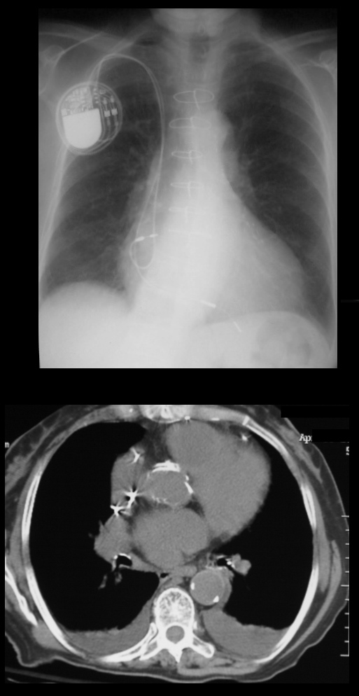
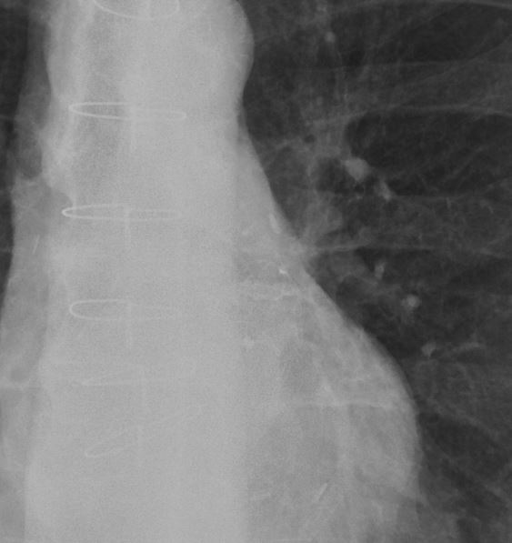
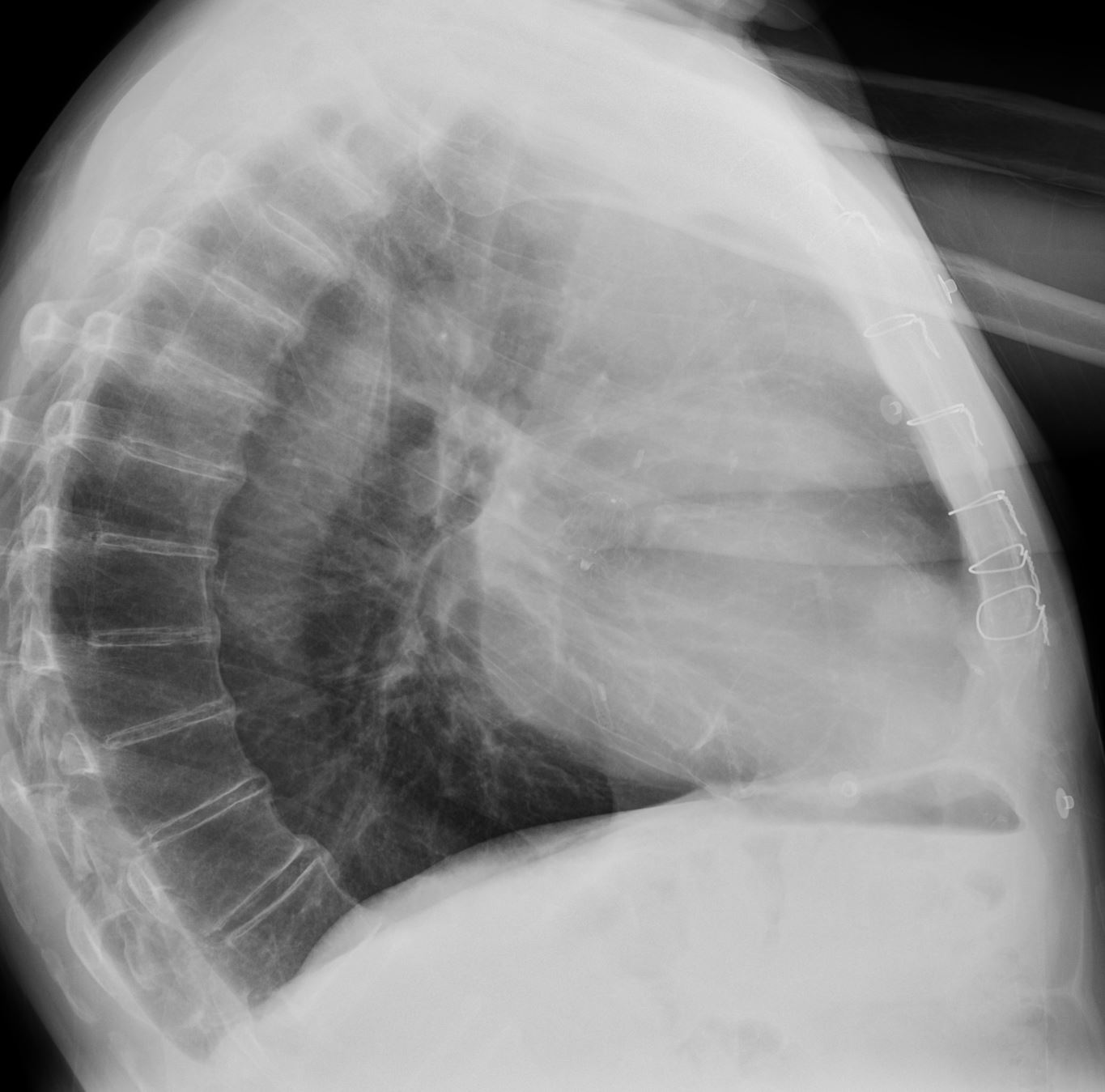
CXR shows a biventricular pacemaker with one wire in the atrial appendage and the second in the apex of the right ventricle.
The CT scan shows the right atrial appendage lead
Ashley Davidoff MD TheCommonVein.net
28767b
Epicardial Leads
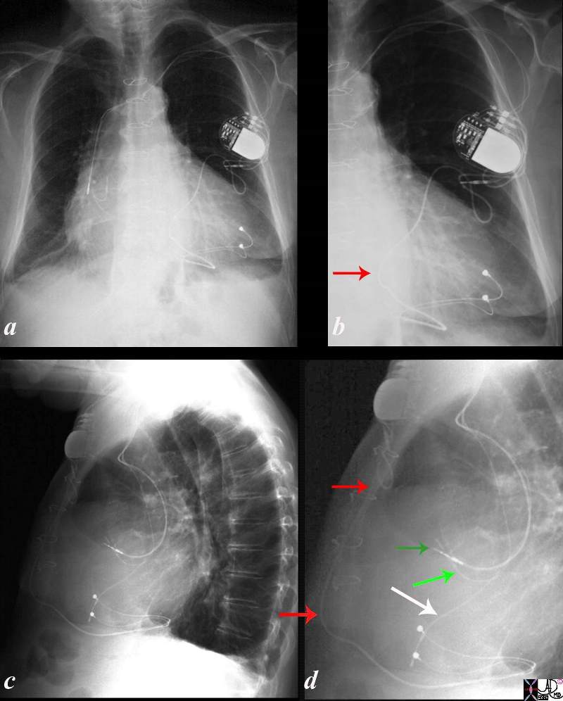
CXR shows a biventricular pacemaker with two wires in the atrial appendage (green arrows) and the second in the apex of the right ventricle (white arrow).
In addition, there is an epicardial lead arising from the battery pack (red arrow). Temporary epicardial leads are frequently placed during cardiac surgery and are then removed in the post op period. In this instance they were probably placed due to failed pacing from the biventricular intracardiac leads
Ashley Davidoff MD TheCommonVein.net
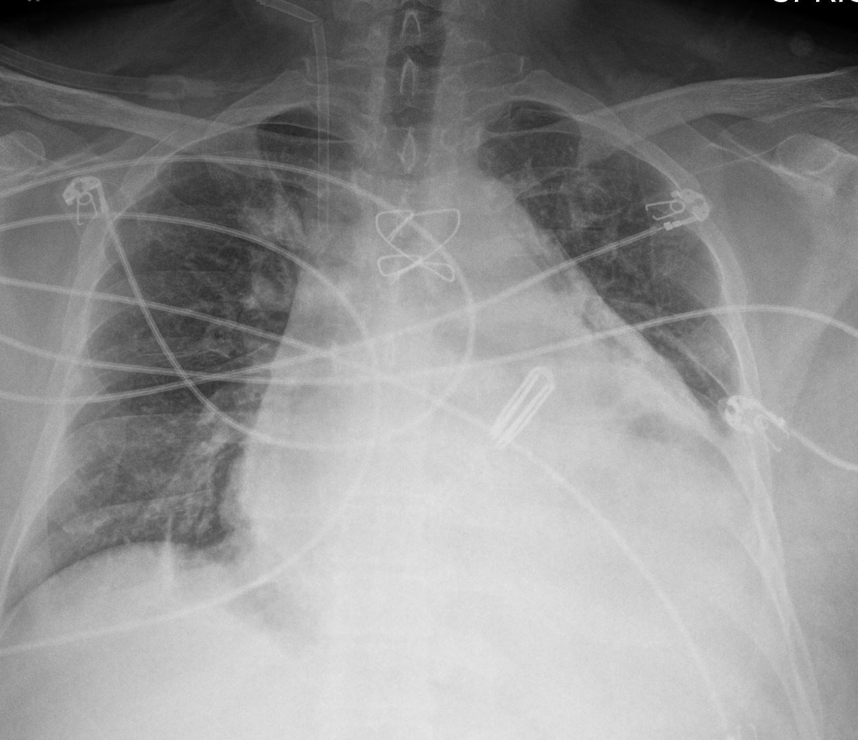
Atrial Appendage Clip
Ashley Davidoff MD TheCommonVein.net
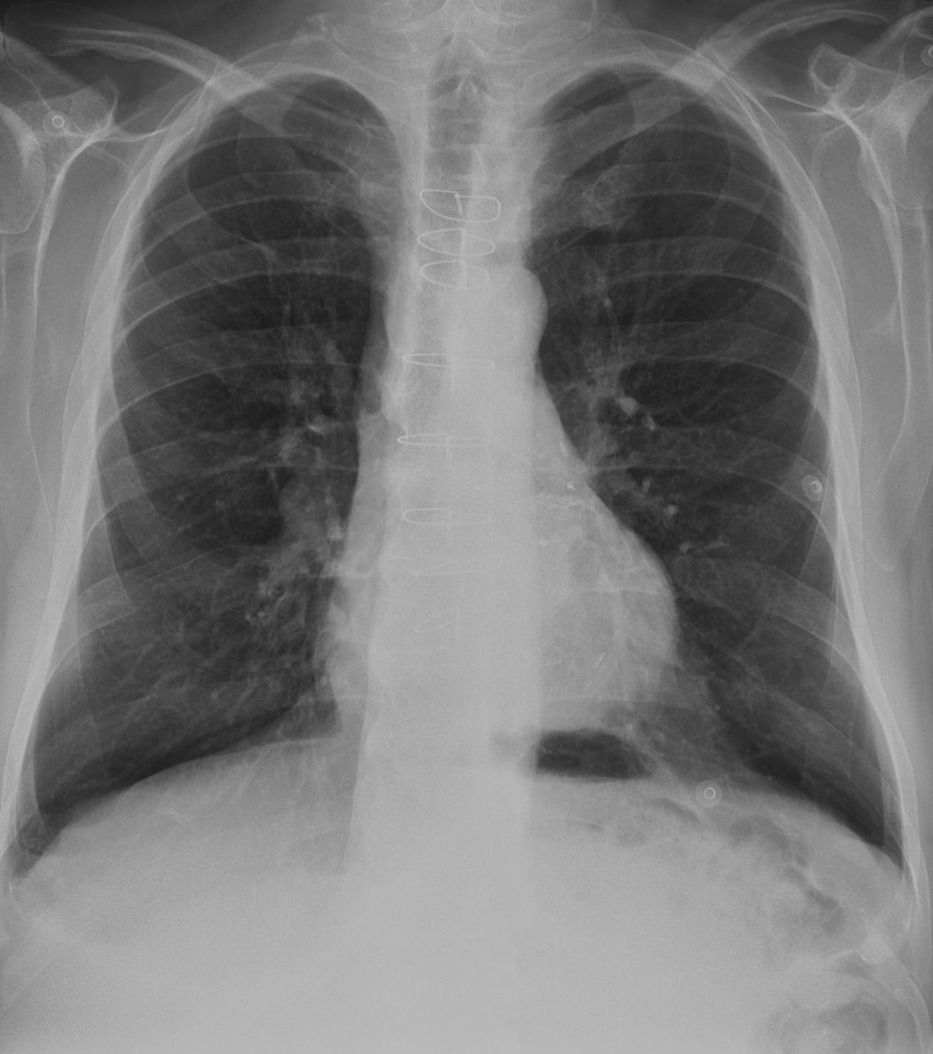
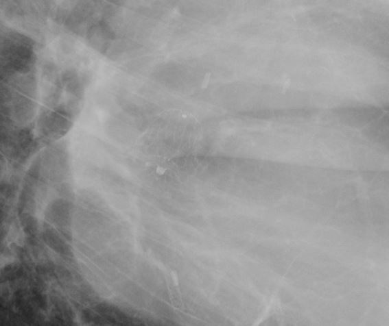
Watchman Device
68 year old male with Watchman Device Ashley DAbvidoff TheCommonVein.net device CXR 68M 001
Links and References
- Radiography of Cardiac Conduction Devices: A Comprehensive Review. by Amanda L. Aguilera et al. RadioGraphics 2011; 31:1669-1682
- K Burney, N Thayur, SA Husain, RP Martin, P Wilde. Imaging of implants on chest radiographs: a radiological perspective. Clin Radiol 2007;62:204-212.
- Patent foramen ovale closure or medical therapy for secondary prevention of cryptogenic stroke An update meta-analysis of randomized controlled trialsby Yingxu Ma et al
Medicine (Baltimore). 2018 Aug; 97(34): e11965. - Radiology Assistant Cardiac Devices