
- Pulmonary valve stenosis
- Causes
- various causes, including:
- Congenital heart defects: The most common cause of pulmonary valve stenosis is a congenital heart defect,
- Rheumatic fever: A complication of untreated strep throat, rheumatic fever can cause inflammation and damage to the heart valves, including the pulmonary valve.
- Carcinoid syndrome:
- Endocarditis: An infection of the heart valves, endocarditis can cause damage to the valves, including the pulmonary valve.
- Idiopathic: In some cases, the cause of pulmonary valve stenosis may be unknown.
- Clinical
- Symptoms can include shortness of breath, fatigue, chest pain, fainting, and heart palpitations.
- Imaging
- Echocardiogram:
- X-ray: A chest X-ray m
- Post stenotic Dilatation
- most specifically of the left pulmonary artery
- CT scan:
- Thickening of the PV
- Enlargement of the pulmonary arteries
- Cardiac catheterization:
-
- Mild PS: Pressure gradient less than 36 mmHg
- Moderate PS: Pressure gradient between 36-64 mmHg
- Severe PS: Pressure gradient greater than 64 mmHg
-
- MRI: Magnetic resonance imaging (MRI) can produce detailed images of the heart and pulmonary valve. This test may be used to evaluate the severity of the stenosis and assess the rx
- RX
- Mild cases may not require any treatment, while more severe cases may require surgery to repair or replace the valve.
- Balloon valvuloplasty may be used as a less invasive alternative to surgery.
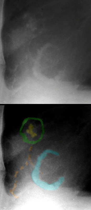
The pulmonary valve is nodular and calcified (yellow) surrounded by a calcified pulmonary annulus (green), associated with a calcified tricuspid annulus (teal), and scattered calcification over or in the right ventricle (orange).
60-year-old man with heart murmur since his teens, but recently became symptomatic with signs of right heart failure characterized ankle edema, ascites and anasarca. Examination confirmed massive ascites and peripheral edema with RV hypertrophy and a coarse ejection systolic murmur in the pulmonic area and absent second heart sound (pulmonic valve closure)
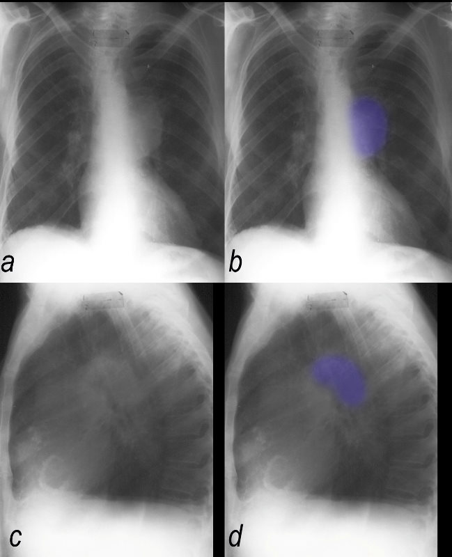
CXR shows aneurysmal dilatation of the left pulmonary artery, decrease pulmonary vasculature, and calcification of the pulmonary valve, pulmonary annulus and tricuspid valve annulus. EKG showed RVH
At catheterization there was a 100 mm Hg gradient across the valve, mean right atrial pressure was 16 mmHg, RV systolic pressure was 120/24 mmHg and PA pressure 25/12 mmHg. Femoral artery O2 sat was 87.5%
Ashley Davidoff MD
60-year-old man with heart murmur since his teens, but recently became symptomatic with signs of right heart failure characterized ankle edema, ascites and anasarca. Examination confirmed massive ascites and peripheral edema with RV hypertrophy and a coarse ejection systolic murmur in the pulmonic area and absent second heart sound (pulmonic valve closure)
CXR shows aneurysmal dilatation of the left pulmonary artery, decrease pulmonary vasculature, and calcification of the pulmonary valve, pulmonary annulus and tricuspid valve annulus. EKG showed RVH
At catherization there was a 100mm Hg gradient across the valve, mean right atrial pressure was 16mmHg, RV systolic pressure was 120/24mmHg and PA pressure 25/12 mmHg. Femoral artery O2 sat was 87.5%
Ashley Davidoff MD
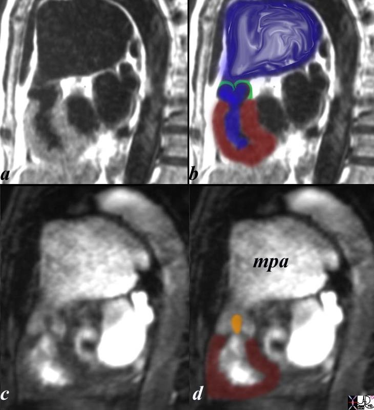
The sagittal view using MRI with black blood (a,b) and white blood(c,d) imaging is from a patient with pulmonary stenosis. The RV (maroon overlay) is hypertrophied, the domed stenotic pulmonary valve (green overlay)is best seen in a and b, and the turbulence through the valve is seen in c, overlaid in orange in d. there is significant post stenotic dilatation of the main pulmonary artery (mpa) inferred by the turbulence in b (blue overlay). code heart cardiac pulmonary valve right ventricle pulmonary artery size enlarged doming stenosis thickened turbulence turbulent flow MRI post stenotic dilatation congenital
Courtesy Ashley Davidoff copyright 2019 15437c03.8s
Radiopedia