
Rare Congenital Abnormality
Unknown Etiology
Resulting in
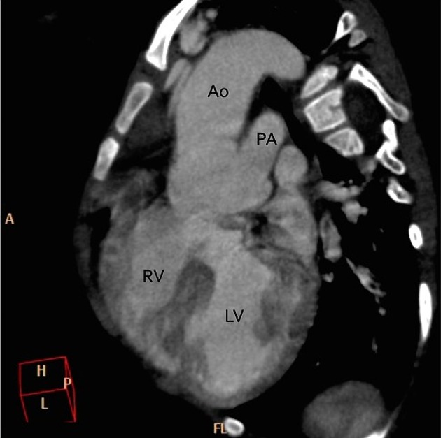
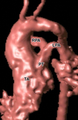
- single artery arising from the two ventricles which gives rise to both the aortic and pulmonary vessels
- abnormal truncal valve
- right sided aortic arch in about 30% of cases (not shown)
- large ventricular septal defect
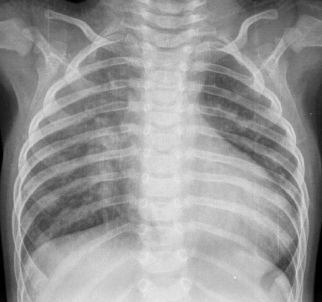
- pulmonary hypertension
- complete mixing occurring at level of the
- right-to-left shunting of blood
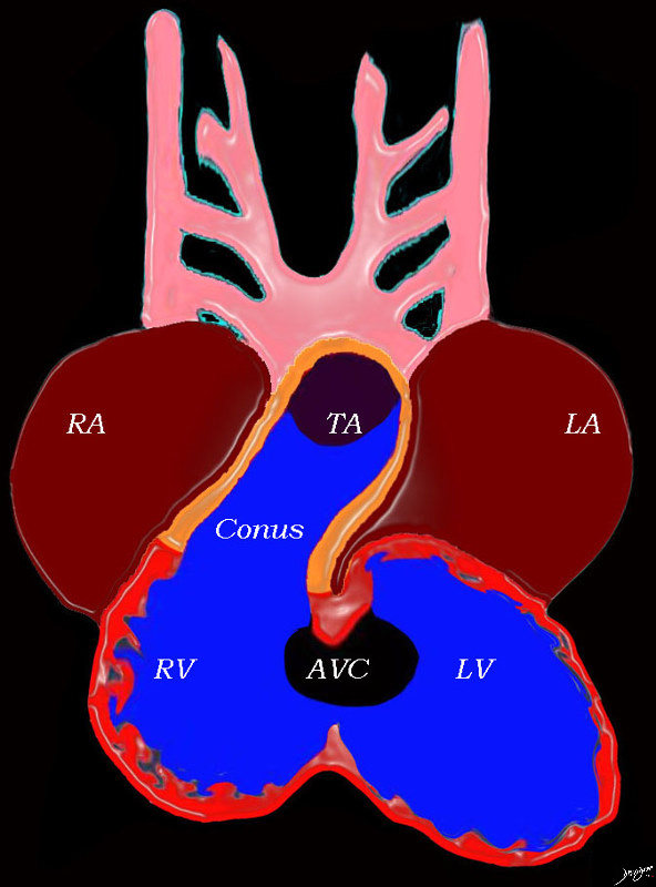
At this stage the heart consists of un-septated atria, ventricles and great vessels, with atria situated posteriorly connected to the ventricles by the AV canal, and the ventricles have a common outlet over the right ventricle to the conus arteriosus, which is connected to the truncus arteriosus
Key words heart cardiac right atrium left atrium right ventricle, left ventricle atrioventricular canal conus arteriosus truncus arteriosus aortic arches RA LA RV LV AVC embryology 01492b04L Ashley Davidoff MD TheCommonVein.net
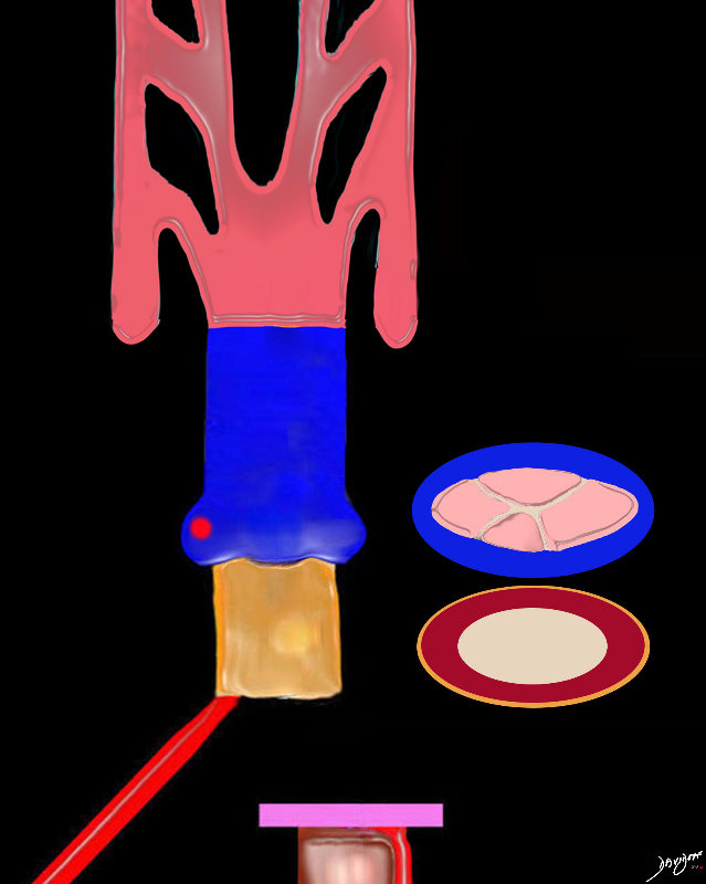
The conus (gold exterior consists of a single muscular tube prior to septation.
Theorigin of the truncus contains 4 truncal cushions which are going to develop into the aortic and pulmonary valve following septation.
Ashley Davidoff MD
TheCommonVein.net
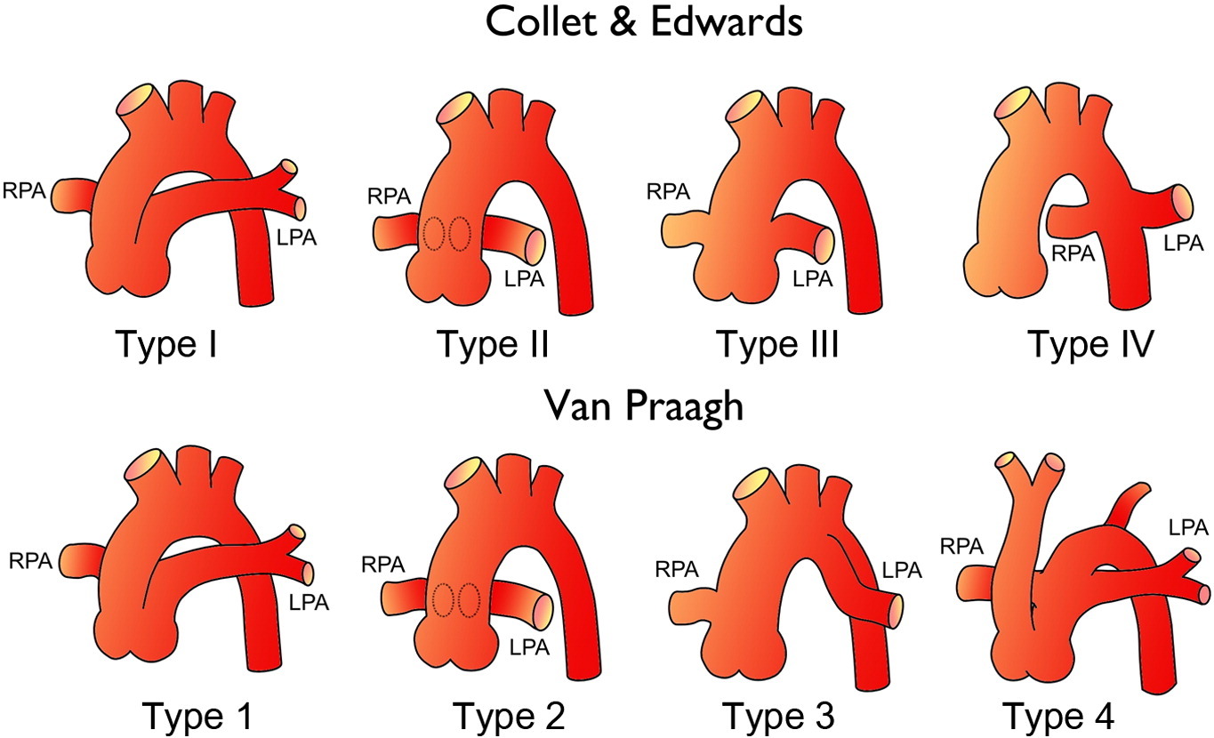
Diagrams show two commonly used systems for classifying truncus arteriosus. Top row: In the Collett and Edwards system, type I truncus arteriosus is characterized by the origin of both pulmonary arteries from a short pulmonary trunk; type II is characterized by the separate origin of the pulmonary arteries from the posterior aspect of the truncus; type III is characterized by the separate origin of the pulmonary arteries from the lateral aspect of the truncus; and type IV is characterized by a pseudotruncus—currently considered a form of pulmonary atresia—with a ventricular septal defect. Bottom row: In the Van Praagh system, types A1 and A2 truncus arteriosus are equivalent to Collett and Edwards types I and II, respectively; type A3 represents atresia of the left or right pulmonary artery, with collateral flow to the ipsilateral lung; and type A4 is characterized by the presence of an associated interrupted aortic arch.
(Frank L et al Cardiovascular MR Imaging of Conotruncal Anomalies Radiographics)
Research Gate
Radiopedia
(Frank L et al Cardiovascular MR Imaging of Conotruncal Anomalies Radiographics)
Classification
Stella and Richard Van Praagh in 1965.[11][12] In this classification scheme, the preceding letter (“A” or “B”) refers to the presence or absence, respectively, of a ventricular septal defect. Type B common arterial trunk is extremely rare; so below, only Type A is considered:
- Type A1: The branch pulmonary arteries arise from a single “main pulmonary artery” arising from the lateral surface of the common trunk (Collett & Edwards Type I)
- Type A2: The branch pulmonary arteries arise separately off the common trunk (includes both Collett & Edwards Types II and III).
- Type A3: One branch pulmonary artery arises off the common trunk, and one branch pulmonary artery is isolated, arising from a patent ductus arteriosus.
- Type A4: Common arterial trunk in association with interrupted aortic arch.
References
- TCV
- Conotruncal Abnormalities
- Embryology
- Anatomy of the Right Ventricle
- Double Outlet Right Ventricle
- Double Outlet Left Ventricle
- Tetralogy of Fallot
- Transposition of the Great Vessels
- Truncus Arteriosus