
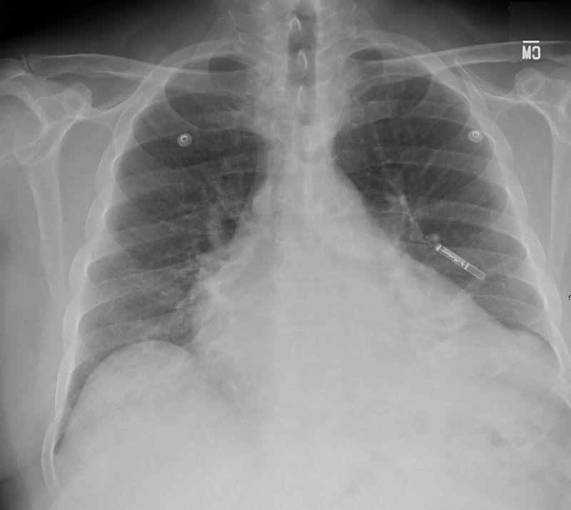
66 year old male presents with dyspnea
Frontal CXR shows LAE and LVE but no evidence of CHF
Note leadless external pacemaker.
Ashley Davidoff MD
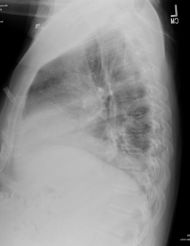
66-year-old male presents with dyspnea
Lateral CXR shows LVE and no evidence of RVE.
Note leadless external pacemaker
Ashley Davidoff MD

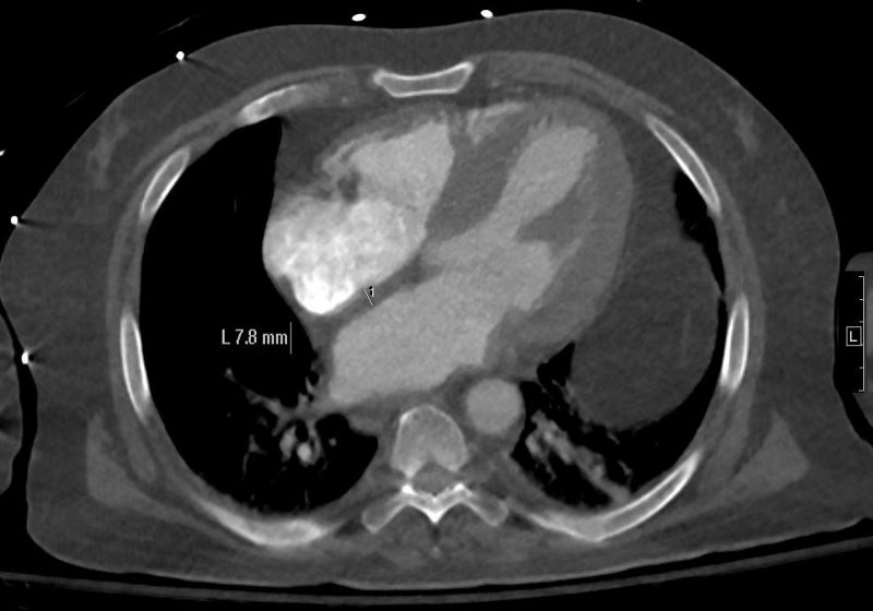
66-year-old male presents with dyspnea
Non gated axial CT through the left atrium at the level of the base of the ascending aorta reveals an A-P dimension of 4.7cms which is slightly enlarged (normal up to about 4.1cms)
Ashley Davidoff MD
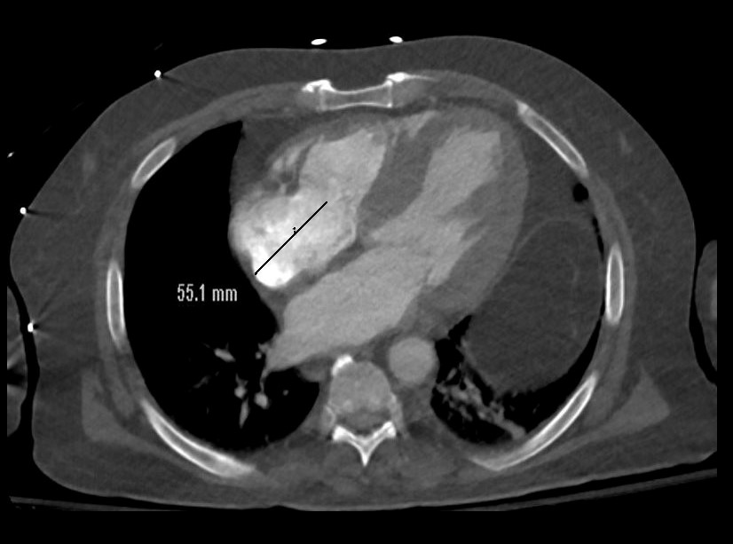
66-year-old male presents with dyspnea
Non gated axial CT through the mitral valve suggests left ventricular hypertrophy. The interatrial septum measures 1.8 mms which in this region is upper limits normal (1.8mms)
Ashley Davidoff MD
66 Male presents with known amyloidosis on MRI and now with increasing dyspnea. CT scan in axial projection shows a right atrium (RA) measuring 5.5cms which upper limits normal to slightly enlarged (normal about 5cms)
Ashley Davidoff MD

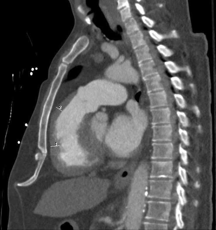
66-year-old male presents with dyspnea
Non gated axial CT through the left atrium at the level of the mitral valve in early diastole reveals a transverse dimension of the RV of 3.3cms (normal up to about 4cms) and a a transverse dimension of the LV of 2.6cms which is small (normal up to about 5cms)
Ashley Davidoff MD

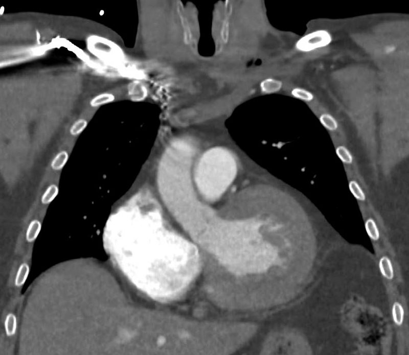
66-year-old male presents with dyspnea
Non gated axial CT through the opening of the mitral valve suggests early diastole confirms concentric hypertrophy. The septum measures 24.1mms while the free wall measures 19.7mms. Upper limits normal is 14mms.
Ashley Davidoff MD
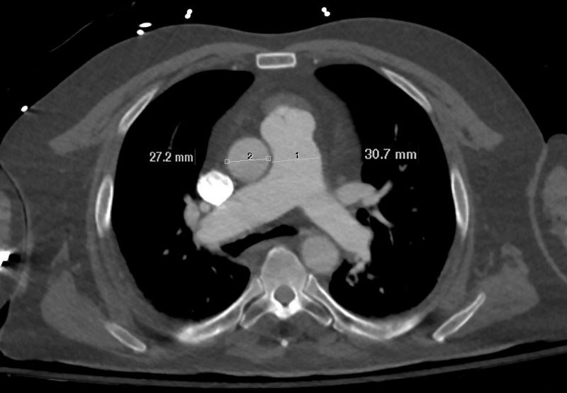
66-year-old male presents with dyspnea
Non gated axial CT through the PA and Aorta at the level of the bifurcation reveals a transverse dimension of the MPA of 3.1cms (normal up to 3cms) and a a transverse dimension of the aorta of 2.7cms(normal up to 3. 5cms)
Ashley Davidoff MD
66-year-old male presents with dyspnea
Non gated coronal CT through the LVOT reveals evidence of LVH
Ashley Davidoff MD
66-year-old male presents with dyspnea
Non gated sagittal CT through the RVOT shows RVH (right ventricular hypertrophy involving both the RV inflow as well as the outflow
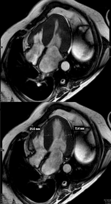
66-year-old male presents with dyspnea
Gated 4 chamber SSFP MRI sequence through the LV during systole (above) and diastole (below) shows concentric hypertrophy. The septal wall measures 25.6mm and the free wall of the RV measures 15.4mm. (normal up to 14mms.)
Ashley Davidoff MD
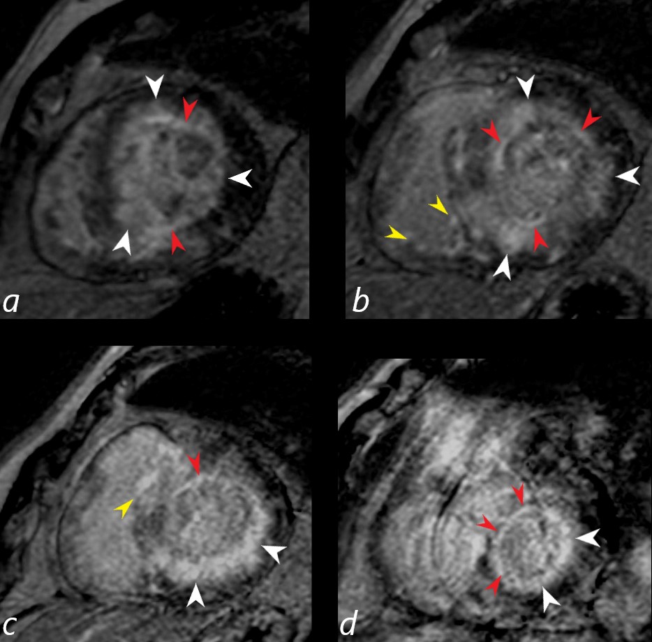
Gated short axis delayed gadolinium sequence through the base LV during diastole and shows subendocardial LGE (red arrowheads in a,b,c, and d, diffuse mid myocardial LGE (white arrowheads) (a,b,c,d) and subepicardial LGE in the RV (yellow arrowheads (b,c)
Ashley Davidoff MD
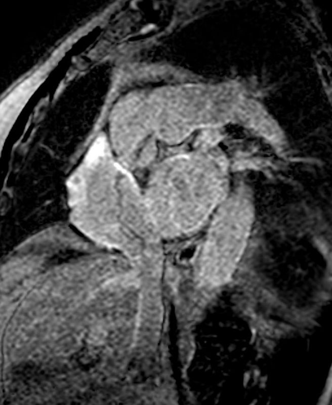
Gated short axis delayed gadolinium sequence through the atria shows LGE in the walls of both atria
Ashley Davidoff MD
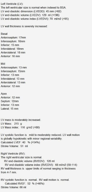
Report of MRI
Report of MRI
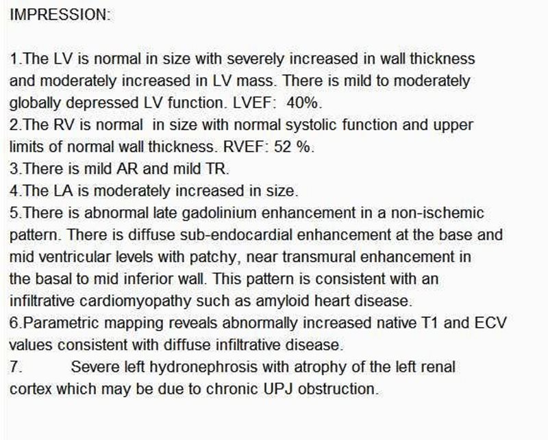
Report of MRI
- 006H Cardiac Amyloidosis
- 032H CAD Amyloid Pericardium Question
- 038Lu Amyloidosis Hilar Lymph Nodes Pericardium CAD
- 66Lu Amyloid nodules alveolar septal and bronchiole and MAC