
MRI
MRI is not a routine method for the evaluation of heart size due to the accurate capabilities of echocardiography which is safe, inexpensive and accurate. However there are many advantages that gated cardiac MRI offers in the evaluation of the heart.
Buzz
- Overall volume
- Shapes
- Linear Size
- Wall thickness
Linear Measurements

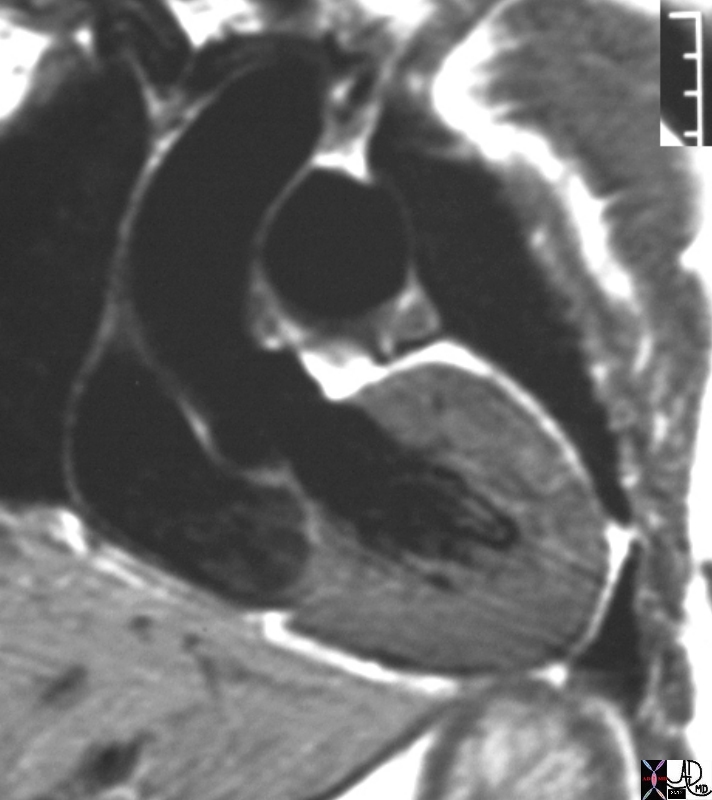
This is the MRI of a 19-year-old male who presented with syncope and the study was performed to identify a possible arrhythmogenic focus
White blood imaging using 4 chamber view shows a normal sized heart in diastole
The septum of the LV in diastole is 8mm, and the free wall is 8mms (upper limits normal is 1.2 -1.4cms. The transverse measurement of the LV cavity is 4.9cms with upper limits normal being about 5 cms.
Ashley Davidoff MD
This is the MRI of a 19-year-old male who presented with syncope and the study was performed to identify a possible arrhythmogenic focus
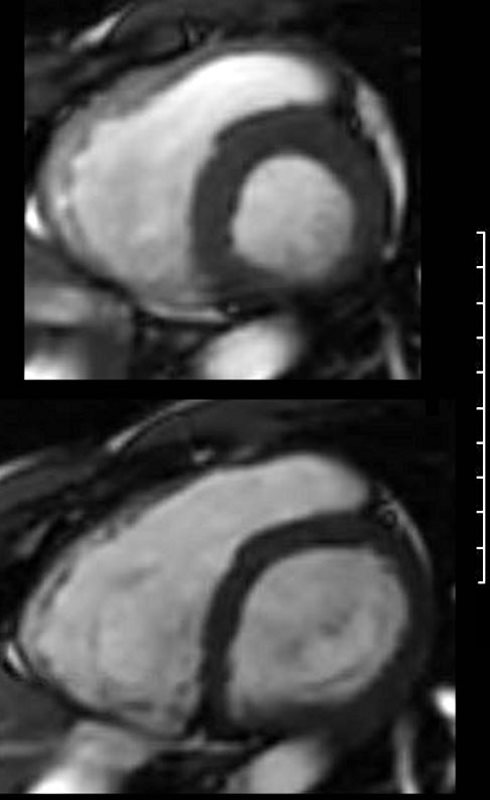
White blood imaging using short axis view shows a normal sized heart in systole (above) and diastole (below). The left and right ventricles show normal wall thicknesses and the volume of the chambers in systole are about 2/3 the volume in diastole (normal). There is no obvious dyskinetic segment of the RVOT.
Ashley Davidoff MD
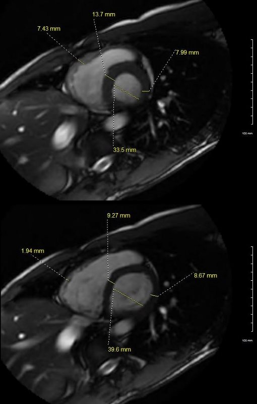
This is the MRI of a 19-year-old male who presented with syncope and the study was performed to identify a possible arrhythmogenic focus.
White blood imaging using short axis view shows a normal sized heart in systole (above) and diastole (below). The transverse dimension of the LV is 4cms in diastole which is normal. The septum of the LV in diastole (lower image) is less than 9.2mms, and the free wall is 8.7 mms (upper limits normal is 1.2cms. The wall of the RV is barely seen in diastole and measures about. In systole the residual volume of the RV is about 1/3 the diastolic volume indicating an approximate ejection of 2/3 = 66% ejection fraction (EF). Similarly, at peak LV systole the residual volume of the LV is about 1/3 the diastolic volume indicating an approximate ejection of 2/3 = 66% ejection fraction (EF).
Ashley Davidoff MD
3 Chamber LVOT
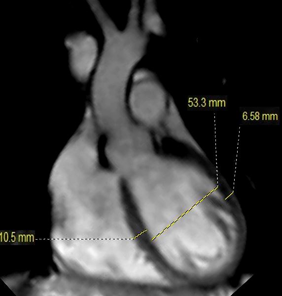
This is the MRI of a 19-year-old male who presented with syncope and the study was performed to identify a possible arrhythmogenic focus
White blood imaging of the LVOT view shows a normal sized ovoid LV in diastole. The septal thickness in diastole is 10.5mms, and bulges toward the RV while the free wall dimension is 6.6 mms. The LV cavity measures 5.3cms which is upper limits normal.
Ashley Davidoff MD

In the gated MRI systole and diastole can be differentiated and so standard measurement to thickness, and volume can be applied for evaluation of size.
This MRI series demonstrates the heart in systole and diastole. Image 1 demonstrates ventricular systole. The atrial chambers are full, the A-V valves are closed and the ventricular chambers are contracted. Image 3 is a color overlay of the closed A-V valves of image 1. Image 2 demonstrates ventricular diastole. The atrial chambers are emptying, the A-V valves are open and the ventricular chambers are full. Image 4 is a color overlay of the open A-V valves of image 2.
32073 Courtesy Philips Medical Systems
tags 16906.800 heart LV left ventricle aorta coarctation left ventricular hypertrophy fx LVH concentric hypertrophy dx coarctation imaging radiology T1 weighted MRI Courtesy Ashley Davidoff MD
Linear Dimensions
- RA
- Systole
- male
- female
- Diastole
- male
- female
- Systole
- LA
-
- Systole
- male 3.3cms
- female 3 cms
- Diastole
- male 5cms
- female 4.5cms
- Systole
-
- RV transverse
- Systole
- male 2.8
- female 2
- Diastole
- male 4
- female 3
- Systole
- LV
- Systole
- male 3.3cms
- female 3 cms
- Diastole
- male 5cms
- female 4.5cms
- Systole
RV LV 4 5
The average transverse diameters of both ventricles, which are important for estimating cardiac dilation, are: female / male left ventricle diastolic 45.2 ± 3.4 / 51.6 ± 4.6 mm, systolic 30.5 ± 3.5 / 33.8 ± 3, 6 mm,
right ventricle diastolic 30.7 ± 3.8 / 37.1 ± 5.9 mm and
systolic 22.3 ± 3.8 / 28.1 ± 4.4 mm.
The septum thickness relevant for left ventricular hypertrophy measures diastolic 8.0 ± 1.0 / 9.9 ± 1.2 mm and
systolic 10.9 ± 1.4 / 13.6 ± 1.9 mm in the short axis of female / male subjects ,
Links and References
Alfakih et al Normal human left and right ventricular dimensions for MRI as assessed by turbo gradient echo and steady‐state free precession imaging sequences JMRI Volume17, Issue3 March 2003 Pages 323-329
Hergan, K et al Normal Cardiac Diameters in Cine-MRI of the Heart in Heart RöFo 2004; 176(11): 1599 – 1606