Copyright 2009
Definition
The left atrium , is a thin walled chamber of the heart which pumps blood into the left ventricle for ejection into the systemic circulation1. Structurally it is smaller than the RA but thicker measuring approximately 3 mms and consists, of three parts (auricle, vestibule and venous compartment). Functionally it receives oxygenated blood from the pulmonary veins and pumps it forward through the mitral valve into the LV to supply oxygenated blood to the tissues of the body.
Diseases of the left atrium are usually secondary to mitral valve or LV failure and present as atrial arrhythmias consequent to LA dilation.
The diagnosis of LA enlargement is suspected in patients with long standing LV failure or mitral stenosis presenting with palpitations, hoarseness of voice or CVA. Chest radiography may reveal a straightening of the left heart border, splaying of the carinal angle – or double density of the right heart border.EKG3 shows P wave duration > 0.12s, notched P wave in limb leads with the inter-peak duration > 0.04s and terminal P negativity in lead V1 . 2D echocardiography is very useful is assessing the LA size and the presence of mural thrombus which are common after LA enlargement.
Medical therapy is used to treat the heart failure, prevent the formation of clots and control the arrhythmias. Surgery and catheter ablations may be required in cases of mitral valve disease or for non responsive atrial arrhythmias.

Ashley Davidoff
TheCommonVein.net
77612b.3kb07.8s
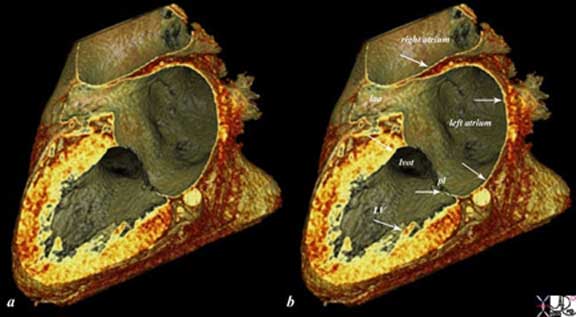
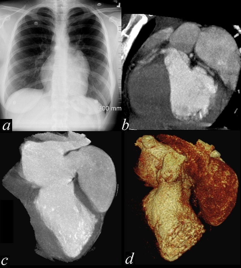
The reconstructed CTscan shows the thin endocardium (white arrows throughout the heart – which is in fact one continuous sheet of tissue connected across the whole circulatory system. The thin white layer is seen in the left atrium, left atrial appendage (laa) over the posterior leaflet of the mitral valve (pl), in the left ventricle (LV) and in the right atrium. The surface of the left ventricle, left ventricular outflow tract (lvot) and right atrium are also lined by the endothelium.. Ashley Davidoff
TheCommonVein.net
47824c02.8s
Principles
Structural Considerations:
The left atrium is relatively featureless internally and possesses a venous component, a vestibule and an appendage, and shares the septum with the RA. The four valveless pulmonary veins, open into the upper part of the posterior surface of the left atrium, two on either side of its middle line. The two left veins frequently end by a common opening.
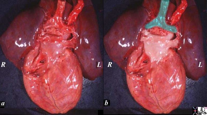
The specimen of heart and lungs is examined from posteriorly with the left atrium and 4 pulmonary veins. (overlaid in pearl white) The trachea and bronchi are intact (green). The left atrium lies just below in the angle of the carina.
Ashley Davidoff
TheCommonVein.net
08376c02.82s
Venous component : Dominant portion of the left atrium and receives one of the four pulmonary veins at its corners and is directly confluent with the body.
Vestibule: Surrounding the mitral orifice is the vestibular component.
Venous component and vestibule are smooth walled as they are derived from absorption and incorporation of the common pulmonary vein into the wall of the left atrium.
Anatomy of the Left Atrium
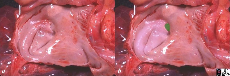
The anatomic specimen has been opened from its posterior aspect revealing a relatively smooth walled inner lining. The left atrial appendage(laa) has been opened revealing fine pectinate muscles. 5 pulmonary veins are seen entering the left atrium, with 2 entering from the left lower vein. The atrial septum with fossa ovalis and foramen ovale are smooth except for the “u” shaped upper border of the septum primum that allows identification of the left atrium. the glistening surface of the endothelium is exemplified in this image.
Ashley Davidoff
TheCommonVein.net
06426c01.81s
Atrial Appendage5: The LAA is a long, tubular, hooked crenulated structure with a narrow junction with the venous component of the atrium. It is derived from the primitive atrium and hence trabeculated, with muscle bars largely running parallel to each other giving a comb-like appearance (hence termed pectinate muscles). However the trabeculations are less pronounced on the left, a useful distinction in some cases of complex congenital heart disease.
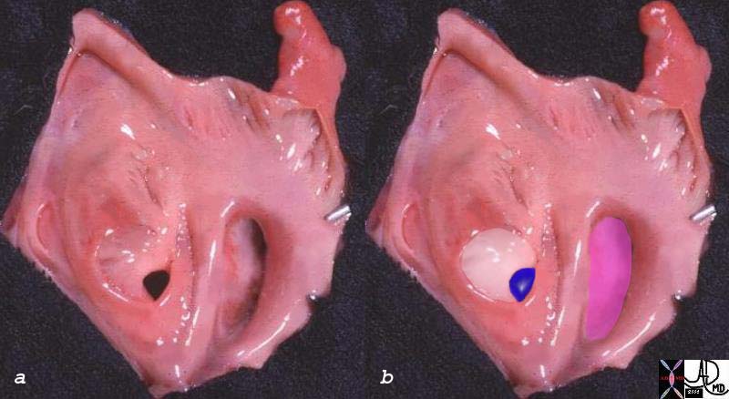
The left atrium does have curves to its inside surface but not to the extent of the right atrium, and it is mostly smooth. The septum primum (white) and foramen ovale (blue) are in the center. The mitral valve is overlaid in pink. Fine pectinate muscles are seen at the mouth of the finger like left atrial appendage.
Ashley Davidoff
TheCommonVein.net
The Left Atrium in Diastole and Systole
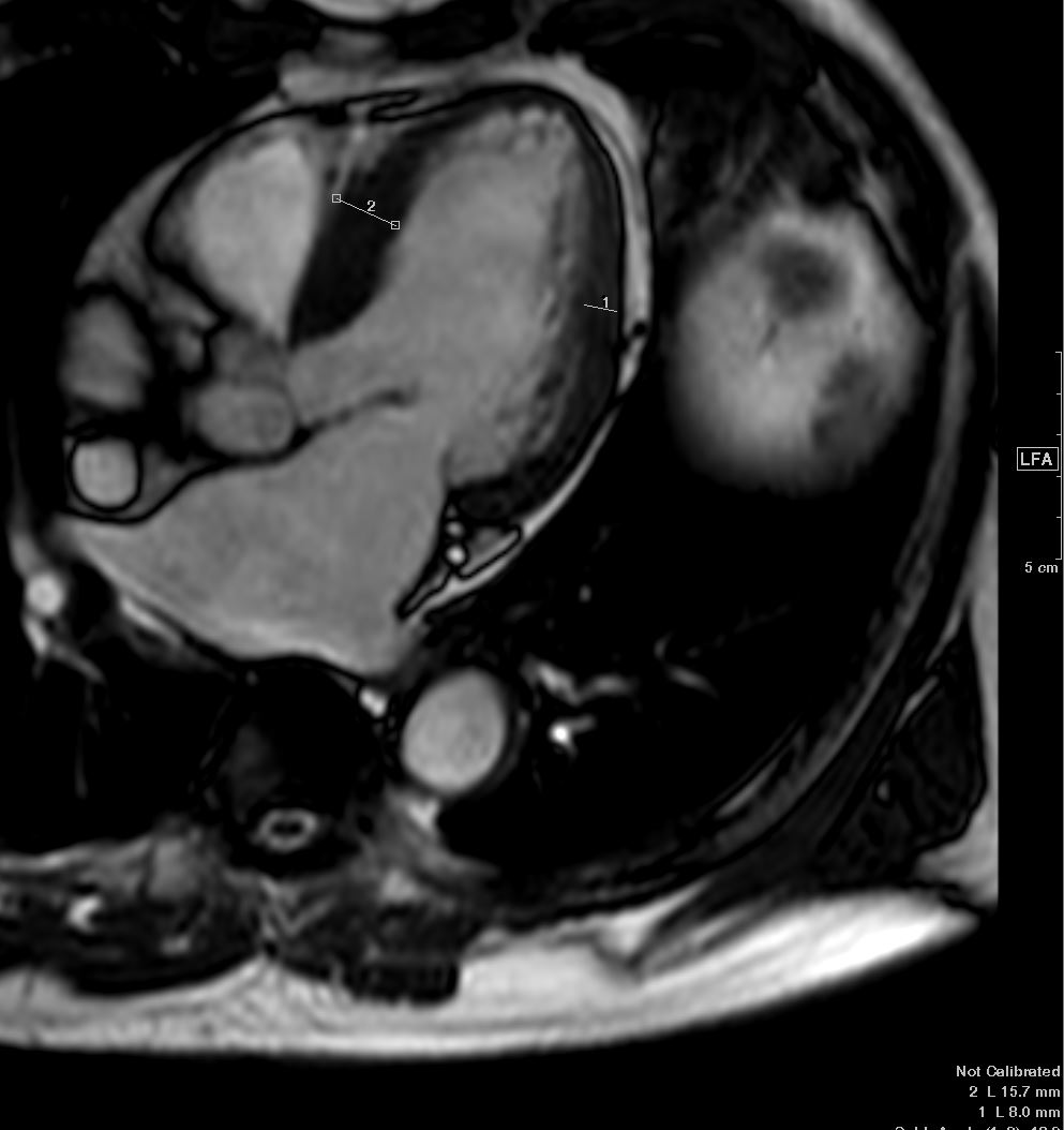
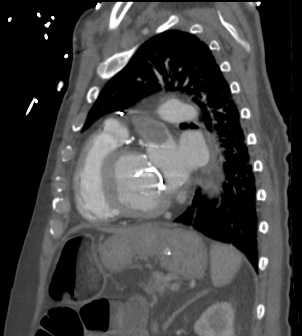
The Mitral Valve is open and therefore we are in mid diastole when the left atrium (LA) is at its emptiest. Note the shape of the left atrium (LA) in diastole and compare it to the shape and volume in systole below
Ashley Davidoff MD TheCommonVein.net wall-thickness-001b

The mitral valve is closed and the walls of the LV have become maximally thickened and therefore we are in peak systole Note the shape of the left atrium (LA) and compare it to the shape and volume in diastole above
Ashley Davidoff MD TheCommonVein.net wall-thickness-002b
Normal LA appendage – MRI
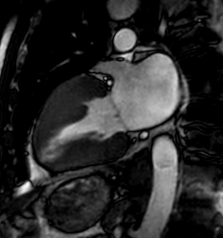
In this 2 chamber view of the LV and LA in systole (mitral valve is closed) shows a normal rotund LA and a normal LAA with concentric thickening of the LV
Ashley Davidoff MD TheCommonVein.net wall-thickness-002

Ashley Davidoff
TheCommonVein.net
Ashley Davidoff
TheCommonVein.net
Ashley Davidoff
TheCommonVein.net
Clinical Considerations:
- Radio-frequency catheter ablation (RFCA) of the distal pulmonary veins and posterior left atrium is increasingly being used to treat paroxysmal or persistent atrial fibrillation4. The esophagus and posterior left atrial wall are in close contact over a wide area and hence increase the potential for complications like esophageal injury and the development of an atrio-esophageal fistula during these procedures.
- Approximately 90% of atrial thrombi in non-rheumatic atrial fibrillation and 60% of such thrombi in patients with rheumatic mitral valve disease (predominantly stenosis) are seen within the LAA5. Therefore prophylaxis against thromboembolism is likely possible by obliterating or amputating the LAA5.
Introduction
The left atrium (LA) is structurally the most boring of the four chambers. It does have a rather strange looking LA appendage but its walls are mostly smooth. It has an interesting position in life in that it lies just beneath the crotch of the carina, and is almost central in the body with a mild bias to the left. Its posterior surface pats the distal esophagus, while its superior surface has the arm of the right pulmonary artery resting upon it.
Size
2-4 cms in its anteroposterior dimension by echocardiography. In cross sectional imaging and echocardiography its size is best assessed by comparing its size in A-P dimension to the size of the aorta.
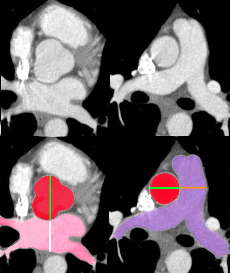
Size Ratios Left Atrium to Aorta Aorta to Pulmonary Artery Ratio |
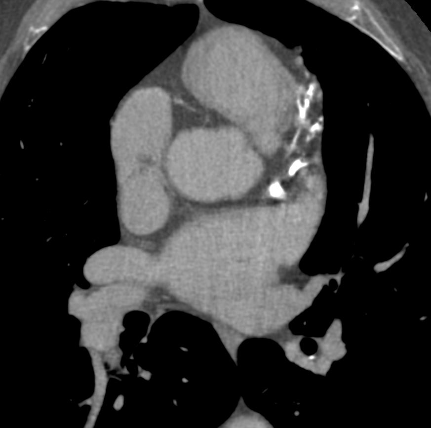
| There are absolute measurements to evaluate cardiovascular structures but imagers find it easiest to compare structures to each other. For the left atrium the A-P dimension of the LA (white line) should be about the same as the diameter of the aorta or the region of the aortic valve (slightly larger). The ascending aorta should have about the same diameter as the main pulmonary artery.
34769c07b02 Davidoff MD Courtesy Ashley Davidoff copyright 2009 |
The enlarged left atrium is sometimes evaluated by the carinal angle.
If the carinal angle is more than 100 degrees – correlates with a LA of greater than 5 cms (Taskin et al)

3D Mitral – Anterior leaflet acting as the boundaries of Inflow |
| 47824 heart cardiac ascending aorta shape aortic tuck mitral valve to aortic fibrous continuity AV MV normal anatomy CTscan Davidoff MD |
|
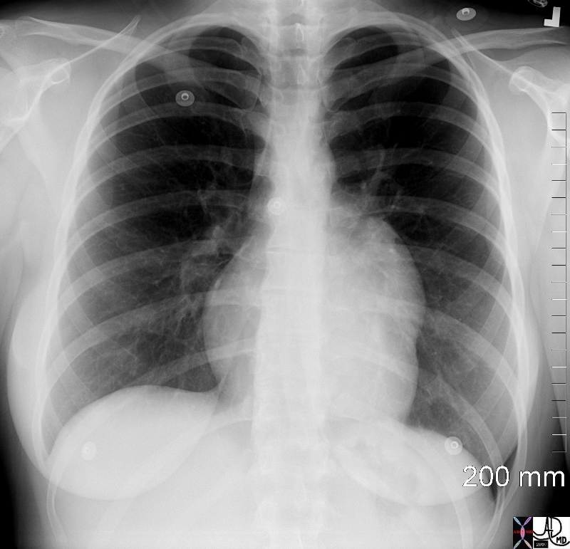
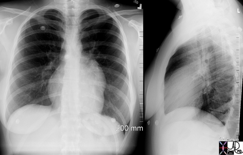
Isolated Left Atrial Enlargement |
|
This plain chest X-ray shows the classical features of an enlarged left atrium characterized by a double density on the right side of the heart (1), splaying of the carina (2), elevation and narrowing of the left main stem bronchus (3), prominent left atrial appendage (4), posterior displacement of the left main stem bronchus (5) as seen as the lateral examination and posterior bulging of the left atrium (6). These findings are consistent with mitral stenosis likely of rheumatic origin Courtesy Ashley Davidoff MD copyright 2009 all rights reserved 15410c02.8s |

Shape
The shape of the left atrium is described based on the projection. In the axial plane it has a more or less rectangular shape, with flattened anterior and posterior borders and rounded lateral borders.
|
Rectangular Shape of the Left Atrium |
|
The axial T1 gadolinium enhanced MRI, (a,b) shows a rectangular shaped left atrium overlaid in bright red) and a similarly shaped left atrium on a contrast enhanced MRI (c,d). The anterior walls tend to be straight and flat and the lateral walls a little more rounded. The left atrial appendage is shown in salmon pink as a finger like projection off the LA, while the tubular pulmonary veins enter into the LA posteriorly Courtesy of Philips Medical Systems and modified by Ashley Davidoff M.D. 32110c01 |

In the coronal plane it usually incorporates the pulmonary veins and looks like the head of a bull.
|
Pulmonary Veins |
|
In the coronal plane the pulmonary veins are usually incorporated into the image and assumes a bull like appearance 34763 Davidoff MD copyright 2009 |
Position
The left atrium is the most posterior of the heart structures with its most immediate posterior relation being the esophagus and then the spine and and aorta. It is the most central of all the chambers but sometimes lies just left of midlene. On its anterior border and to its right is the SVC and RA and to its left and anterior is the coronary artery. Superiorly the right main pulmonary artery branch runs and above that is the carina. Inferiorly the esophagus continues as a posterior and left sided relation, and the base of the heart lies anteriorly and inferiorly.
|
The Position of the Left Atrium |
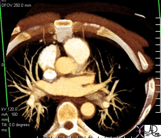
|
This cross sectional CT image shows the pulmonary veins entering the left atrium. It shows its posterior relations that include the aorta and spine, anterior and rightward relation the SVC, the aorta, left coronary artery on the anterior and left aspect Courtesy Ashley Davidoff MD 31236 copyright 2009 |
|
Orientation of the left Atrium in the Coronal Plane |
|
The left atrium is oriented from its central position to the mitral valve which is more anterior and more leftward. 34777.8s all rights reserved copyright 2009 Courtesy Ashley Davidoff cardiac |

|
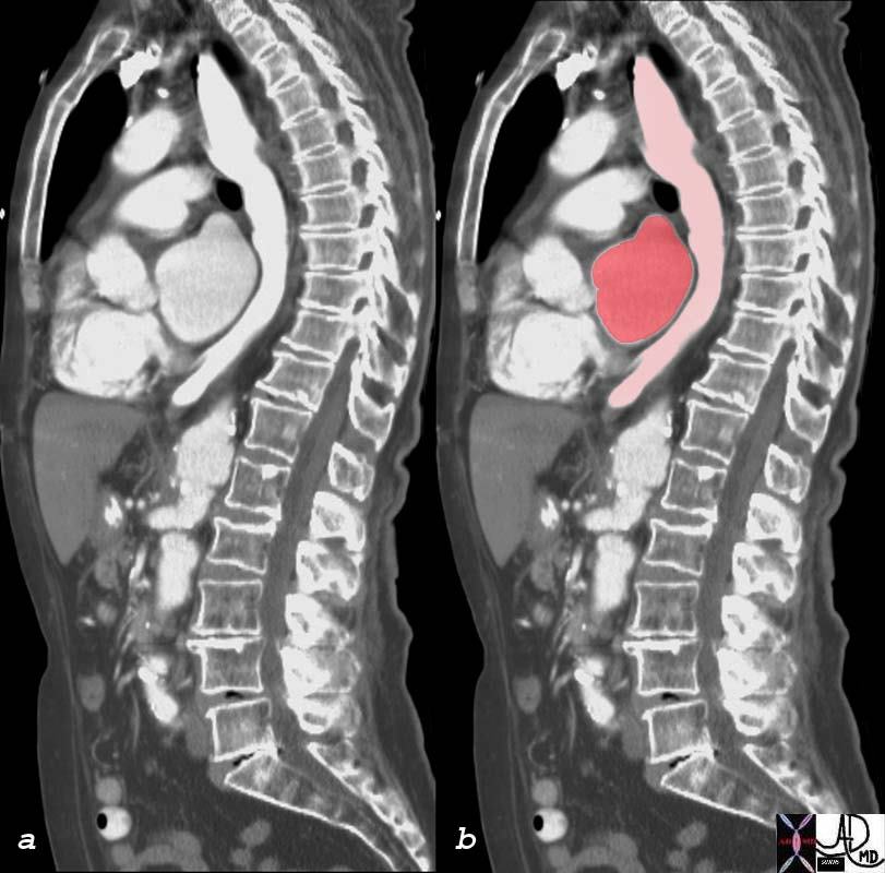
Position in Relation to the Esophagus |
| 75834c01 esophagus left atrium relationship contrast reflux up esophagus (pink) enlarged left atrium impinging on the esophagus (red) CTscan Courtesy Ashley DAvidoff MDm |
Parts
There are not many parts to the left atrium. the most fascinating structure is the left atrial appendage. It has an unusual shape, with irregular fingerlike projections off its upper and lower borders.
|
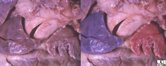
Atrial Appendages |
|
This anatomical specimen shows the right and the left atrial appendages. The right atrial appendage (blue overlay) has a characteristic Snoopy’s nose shape, while the left (red overlay) is narrower, and with multiple frond-like features better appreciated in the next image. Courtesy of Ashley Davidoff M.D. 32103 copyright 2009 |
|
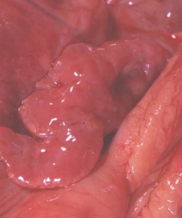
Another Example of the Unusually Shaped LAA |
| A second anatomical specimen of the LA appendage (laa) shows the full extent of the rather odd looking left atrial appendage. Its shape resembles a crooked finger, or the map of South America. Courtesy of Ashley Davidoff M.D. 32104 |
|
Atrial Appendages |
|
CT scan from two different patients highlighting the atrial appendages. The right atrial appendage in a and overlaid in blue in c, is broad and triangular while the left atrial appendage is narrower (b,d) and lobular with a pointed narrowed apex. Copyright 2009 Courtesy Ashley Davidoff MD 34772c01.8s right atrial image from 31232 |

Disorders
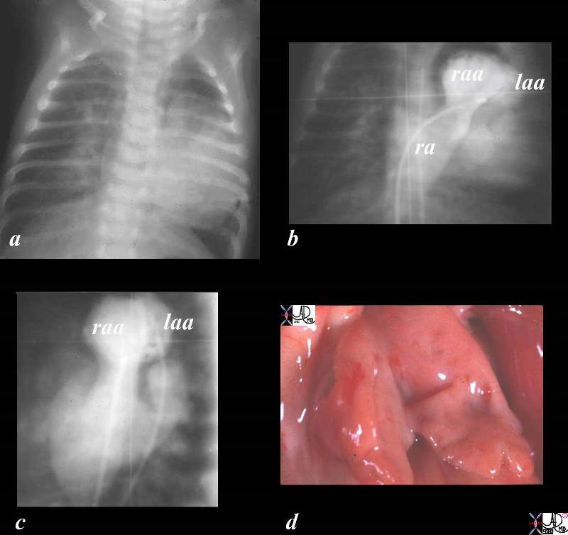
The CXR (a) angio in the P-A projection (b) and lateral projection(c) show positioning of the right atrial appendage on the left side rather than on the right and sits in front of the left atrial appendage. the condition was confirmed at autopsy. the entity is called juxtaposition of the atrial appendages.
Courtesy Ashley Davidoff Copyright 2009 all rights reserved 01865c02.8s
TheCommonVein.net Ashley Davidoff
|
01475 |
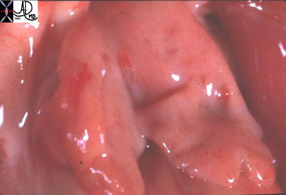
| This gross pathology specimen shows two left atrial appendages pointing leftward in this patient with juxtaposition of the atrial appendages. Courtesy Ashley Davidoff MD 01475 Code CVS heart cardiac congenital LA LAA gross pathology |
Normal Left Atrial Appendage and Aneurysm of LAA |
| 70025d01 heart cardiac left atrium LA left atrial appendage fx enlarged fx aneurysmal dx aneurysm of the left atrial appendage CTscan Davidoff MD |
|
Fetal Ultrasound |
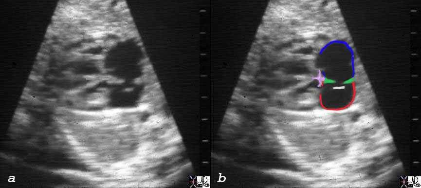
| The fetal ultrasound shows the right atrium in blue, left atrium in red, limbic band in green and the septum primum white, which is open and lies on the left atrial side.
06428c03 heart cardiac atrial septum RA septum secundum superior limbic band inferior limbic band endocardial cushions ventricular septum fossa ovalis fetal echocardiogram LaA left atrium normal anatomy Courtesy Ashley DAvidoff MD copyright 2008 |
|
The Enlarged Left Atrium by CT |
|
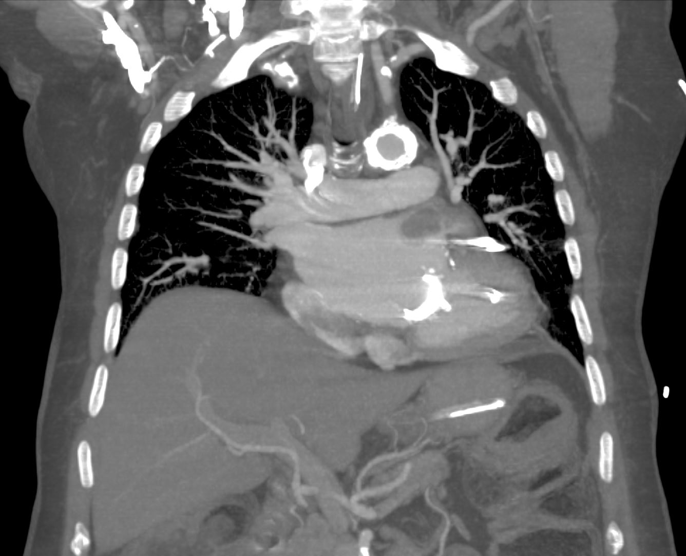
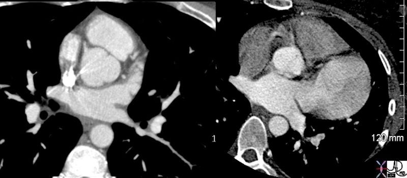
This cross sectional image of the heart through the left atrium and aorta show a left atrium that is greater than 2 x the dimension of the aorta indicating significant left atrial enlargement. Bilateral pleural effusions are present consistent with cardiac failure. Courtesy Ashley Davidoff MD copyright 2009 all rights reserved 16527c02.8s |

|
Compressed Left Atrium |
|
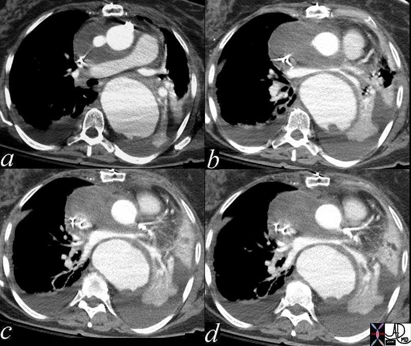
61 year old female post op repair of ascending aortic aneurysm with Bentall procedure with dropping blood pressure and decreasing oxygen saturation. The images show a post op hematoma pushin on the anterior aspect of the left atrium, and an aneurysm of the descending aorta compressing the left atrium from behind. The left atrium is diminutive and looks like a bow tie between the ascending and descending parts of the aorta Davidoff MD copyright 2009 all rights reserved 47752c01 |
Equilibrium Phase Appendage filled with contrast – no LAA thrombus
Ashley Davidoff MD TheCommonVein.net
keywords heart cardiac left atrium LA left atrial appendage fx enlarged fx aneurysmal dx aneurysm of the left atrial appendage CXR plain film chest Ashley Davidoff MD TheCommonVein.net 70025b01
keywords heart cardiac left atrium LA left atrial appendage fx enlarged fx aneurysmal dx aneurysm of the left atrial appendage CXR plain film chest Ashley Davidoff MD TheCommonVein.net 70025c02