
Mild Congestive Cardiomyopathy
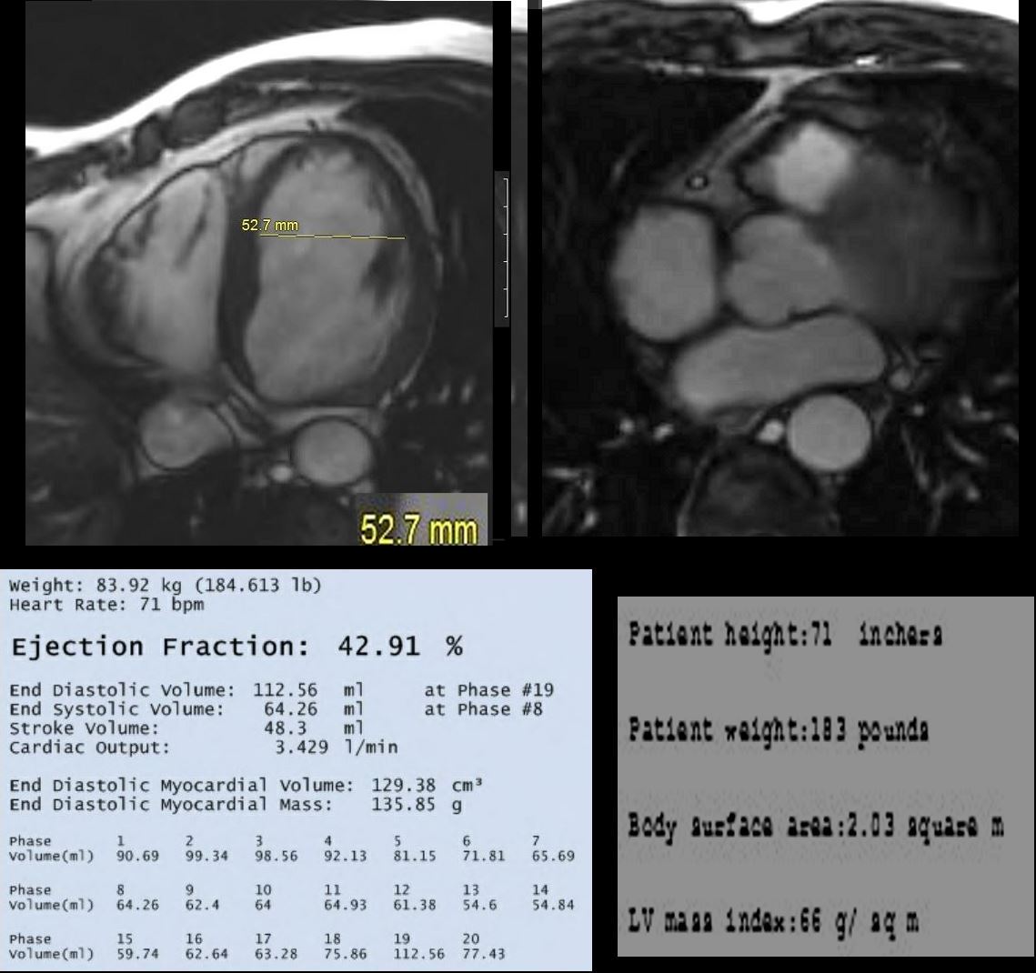
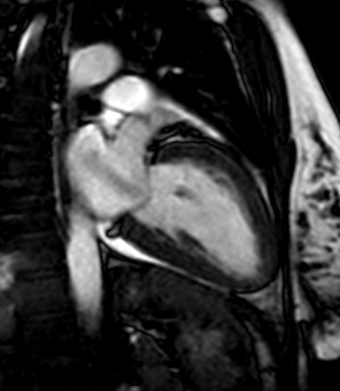
Axial MRI through mid ventricle using white blood imaging algorithm, in a 66 year old male with mild dyspnea, shows mild left ventricular (LV) enlargement (LVE) with an internal diameter of 5.3cms (normal 4.5cms) (top left), normal sized left atrium (LA) approximating the size of the normal aorta (Ao) with an ejection fraction of 43% and normal end diastolic volume (112 ccs) and normal LV mass (66gms/sq m)
Ashley Davidoff MD

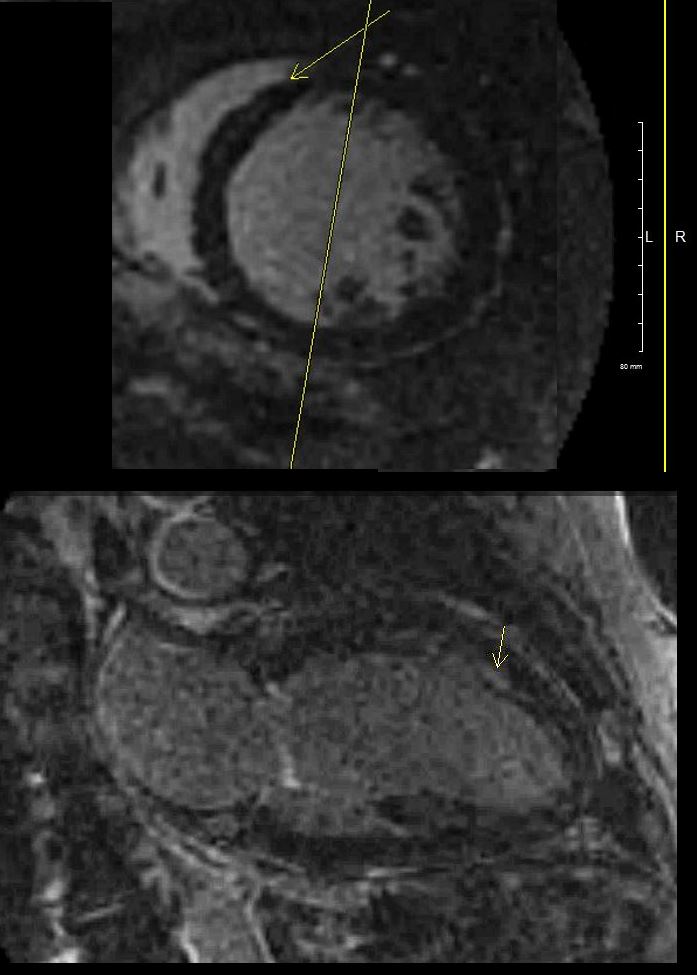
Short axis MRI in a 66 year old male with mild dyspnea, post gadolinium infusion through mid ventricle using white blood imaging algorithm, shows linear enhancement in the mid myocardium of the septum (white arrows in b) consistent with congestive cardiomyopathy. Myocarditis is included in the radiological differential diagnosis.
Ashley Davidoff MD
61 Male with Alcoholic Congestive Cardiomyopathy
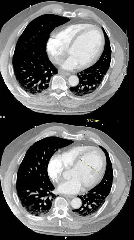
Axial CT through the 4 chambers shows isolated LV dilatation in early diastole with the cavity measuring 6.8cms.
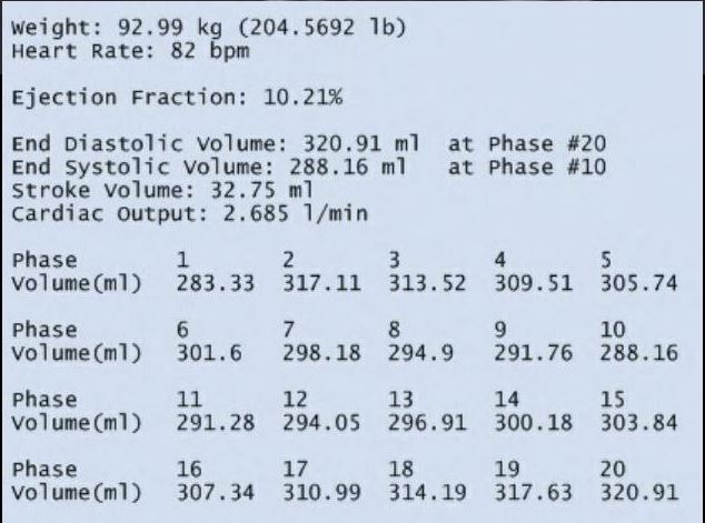
MR in axial plane shows a dilated LV and normal sized LA, RA, and RV. EF was between 10% and 20%
Ashley Davidoff MD
61 male, alcoholic with congestive cardiomyopathy
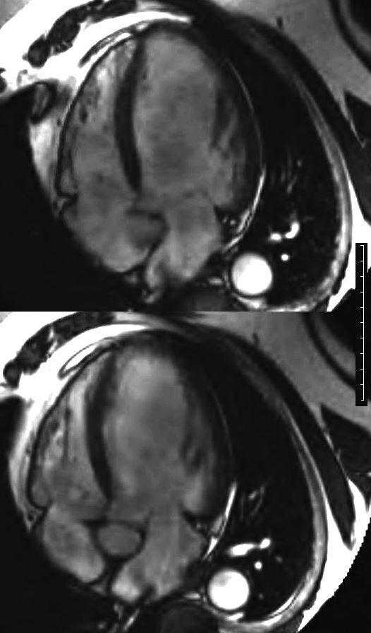
MRI in short axis through the body of the LV in systole (above) and diastole (below)shows a dilated LV without much change in the dimensions of the LV cavity. EF was between 10% and 20%
Ashley Davidoff MD

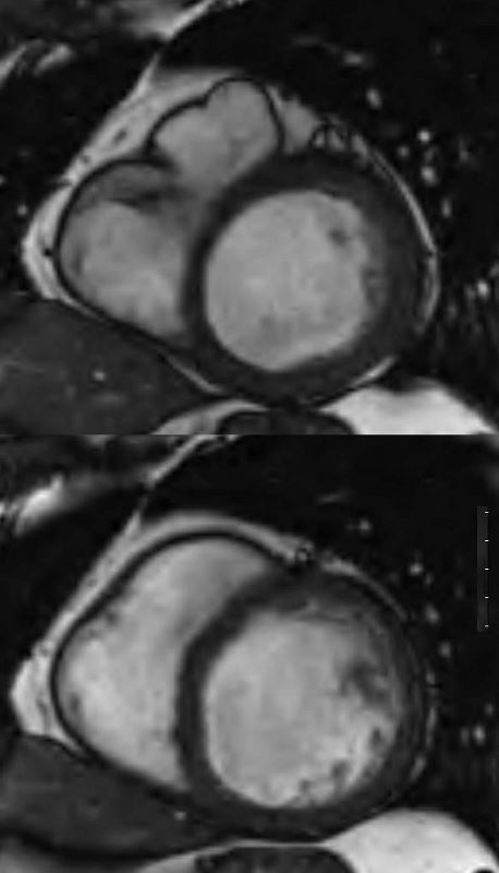
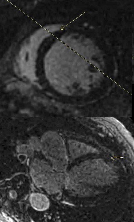
Short axis LGE sequence shows linear mid myocardial LGE in the ventricular septum
Ashley Davidoff MD
CONGESTIVE CARDIOMYOPATHY
61 male, alcoholic, with congestive cardiomyopathy
4 chamber LGE sequence shows linear mid myocardial LGE in the ventricular septum
Ashley Davidoff MD
2 Chamber LGE sequence shows linear mid myocardial LGE in the ventricular septum
Ashley Davidoff MD
40 year old female with SLE and congestive cardiomyopathy in this 2 chambered view shows a jet of mitral regurgitation caused by to ventricular remodeling leading to reduced leaflet coaptation and subsequent valvular insufficiency.
Ashley Davidoff MD TheCommonVein.net mitral-regurgitation-001-MRI
References and Links