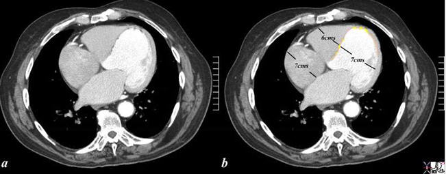
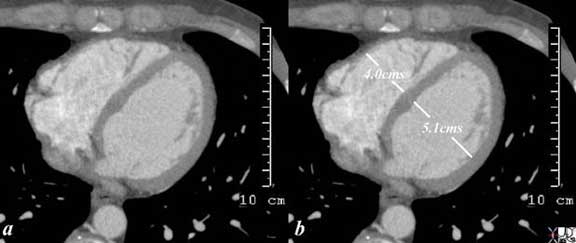
Enlarged Left Ventricle What is the cause of the cardiomegaly? The CT scan is from a 76 year old man in whom the dominant finding is of left ventricular enlargement, characterized by the rotund shape of the ventricle and the increased dimension. The RA and RV are also enlarged based on this image, and LA was enlarged as well suggesting global cardiomegaly consistent with a cardiomyopathy. The clue to the cause of the enlargement is the segmental nature of the disease, characterized by the asymmetry thickness when the free wall thickness is compared to that of the thinning of the septum. In addition, the presence of fat (yellow overlay) in the thinned and probably scarred myocardium, makes ischemic cardiomyopathy the likely diagnosis. Courtesy of: Ashley Davidoff, M.D heart-anatomy-P-047Normal Size of the Right Ventricle and Left Ventricle in the Axial projection During Diastole The axial gated CT scan through the right and left ventricle at end diastole shows the normal size and shape of the right ventricular inflow tract and left ventricle. The right ventricular inflow (underlying the RV measurement) looks smaller than the LV in volume, in this view, since essentially it makes up for the volume in its second “floor” which sits more cranially as the right ventricular outflow tract. The left ventricle only has a single level or floor. Thus in this view the RV looks and measures smaller then the LV. Note also that the apex of the left ventricle protrudes slightly more anteriorly than the RV even though it is the posterior ventricle, because it is the chamber that forms the apex of the heart. The septum also bulges toward the right ventricle due to the higher pressure in the left ventricle. Courtesy of: Ashley Davidoff, MD aka 37758b01c01.8s aka heart anatomy P 040