
Patent Foramen Ovale
The Common Vein Copyright 2008
Ashley Davidoff MD
Definition
A patent foramen ovale is an unsealed septum primum f;ap valve which is normally closed in postnatal life as the left atrial pressures rise and force the septum primum against the superior limbic band. In 70% of patients the foramen seals over permanently but in 30% the valve is functionally closed but not structurally sealed.
This has no functional significance except under circumstances where the right atrial pressures rise above those of the left forcing the flap valve to open and allow unwanted embolic debris to traverse the septum and enter the systemic circulation.
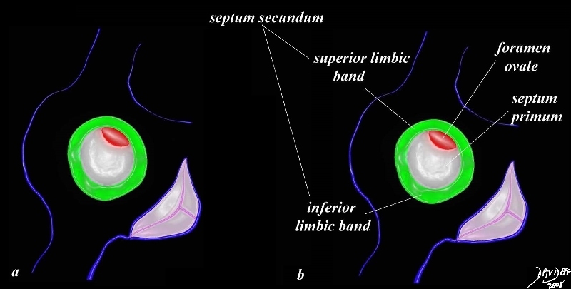
| Fossa Ovalis, Septum Secundum, and Septum Primum
The fossa ovalis is a central structure of the atrial septum that consists of a ring of muscle (green) surrounding a membrane called the septum primum, that has a crescentic defect at the 1 oclock position (red) called the foramen ovale. The superior portion of the muscle is called the superior limbic band, and the inferior aspect is called ythe inferior limbic band. 01656c03.8s Davidoff art copyright 2008 all rights reserved |
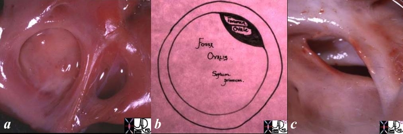
| Fossa Ovalis, Septum Primum, and Foramen Ovale |
| The fossa ovalis is the depressed ring like structure surrounded by a rim of muscle seen in the first image, consisting of the septum primum and foramen ovale seen in the second diagram. An anatomical specimen of a patent foramen ovale is seen in the third image characterized by its position and crescentic shape.01696c01.8s 01671 01670 Davidoff MD copyright 2008 all rights reserved |
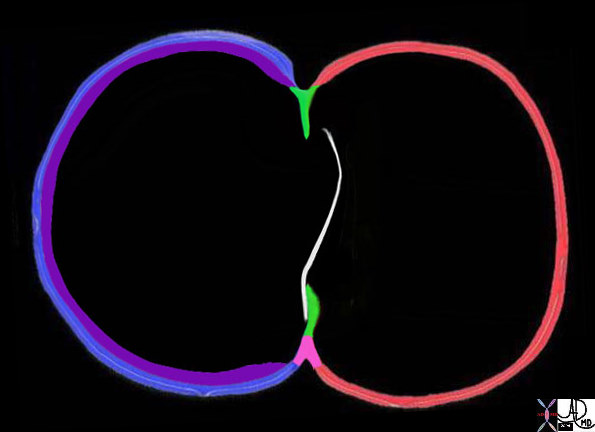
| Atrial Septal Tissues
Sinus venosus = Purple Septum Secundum = Green, AVC = Pink |
| 07538c07 heart cardiac fetal circulation atrial septum septum primum sinus venosus right to left shunt RA right atrium left atrium LA fossa ovalis foramen ovale Davidoff art Courtesy Ashley Davidoff Md copyright 2008 |

| Patent Foramen Ovale |
| The anatomic specimen shows a patent foramen ovale (overlaid in red) on the superior aspect of the septum primum(pink). The image is photgraphed from the left atrium.
heart interatrial septum fossa ovalis septum primum left atrium LA pathology anatomy courtesy Ashley Davidoff MD copyright all rights reserved 01690c01.8s |
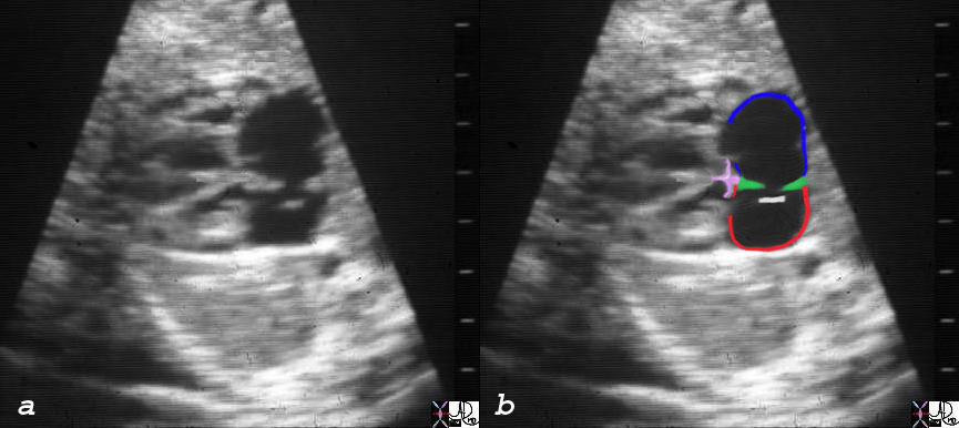
| Fetal Ultrasound |
| 06428c03 heart cardiac atrial septum RA septum secundum superior limbic band inferior limbic band endocardial cushions ventricular septum fossa ovalis fetal echocardiogram LaA left atrium normal anatomy Courtesy Ashley DAvidoff MD copyright 2008 |
Patent Foramen Ovale Paradoxical Embolus
The following series of images is from a remarkable case of a elderly patient who presented to the emergency room with chest pain, shortness of breath, and low oxygen saturations.
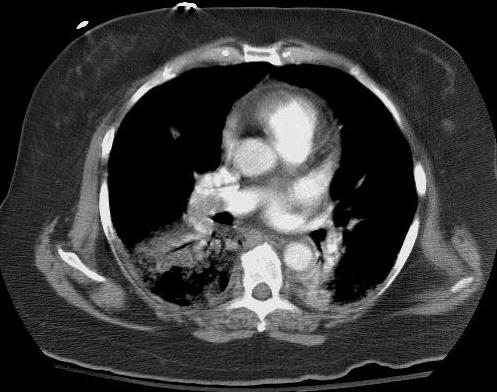
| Pulmonary Embolus |
| Chest ST shows a large filling defect in the right pulmonary artery, and an infiltrate beyond the PE likely representing a pulmonary infarction.
43666 lung fx infiltrate artery lung pulmonary embolus pulmonary atelectasis vs infarction PE PFO imaging radiology CTscan Courtesy Ashley Davidoff MD 43666 43670 43672 43675 43680 43683 5star |
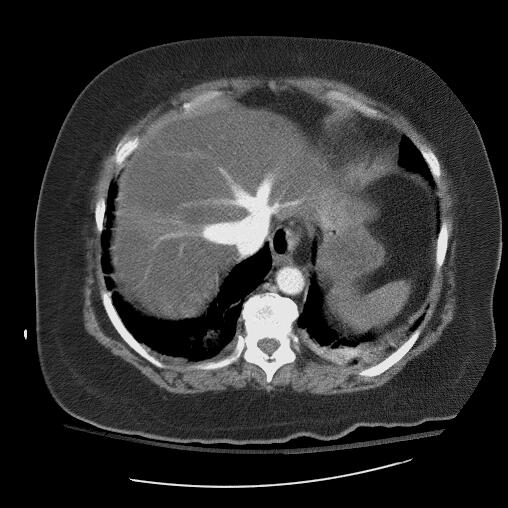
| Tricuspid Regurgitation |
| The last few images of the Chest CT showed reflux of contrast into the hepatic veins suggesting acute tricuspid regurgitation, likely as a result of the sudden increase in pulmonary arterial pressures.
43670 liver hypodense hepatic veins fx enlarged dx tricuspid regurgitation dx PE with secondary pulmonary hypertension and TR 43666 43670 43672 43675 43680 43683 5star |
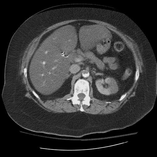
| Renal Infarction |
| The CTscan through the liver and left kidney shows a non perfusing area in the left kidney consistent wit a renal infarction, while a filling defect is suggested in the abdominal aorta.
43672 kidney fx hypodense hypoperfusion fx wedge fx focal swelling enlargement aorta fx filling defect dx pulmonary embolism secondary pulmonary hypertension and TR embolus via PFO dx pulmonary embolus PE imaging radiology CTscan Courtesy Ashley Davidoff MD 43666 43670 43672 43675 43680 43683 5star |
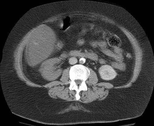
| Renal Embolus -Renal Infarction
Aortic Embolus |
| The CT scan a few cuts lower show a definite filling defect in the aorta and non perfusion of the right lower kidney suggesting infarction of the right kidney as well. Lower down embolic disease was noted in both iliac arteries.
The case reveals the devastation that embolic disease can produce. venous embolism is not uncommon and can be in ite=”self” fatal. In this case the sudden rise in PA pressures caused RA hypertension, TR, right to left shunting through a patent foramen ovale, and systemic emboli to the aorta and iliacs and bilateral real infarctions. 43675 kidney fx hypodense hypoperfusion fx non perfusion kidney infarction aorta fx filling defect crescent dx pulmonary embolism PE secondary pulmonary hypertension and TR embolus via PFO imaging radiology CTscan Courtesy Ashley Davidoff MD 43666 43670 43672 43675 43680 43683 5star |