
66 year old female presents with acute dyspnea
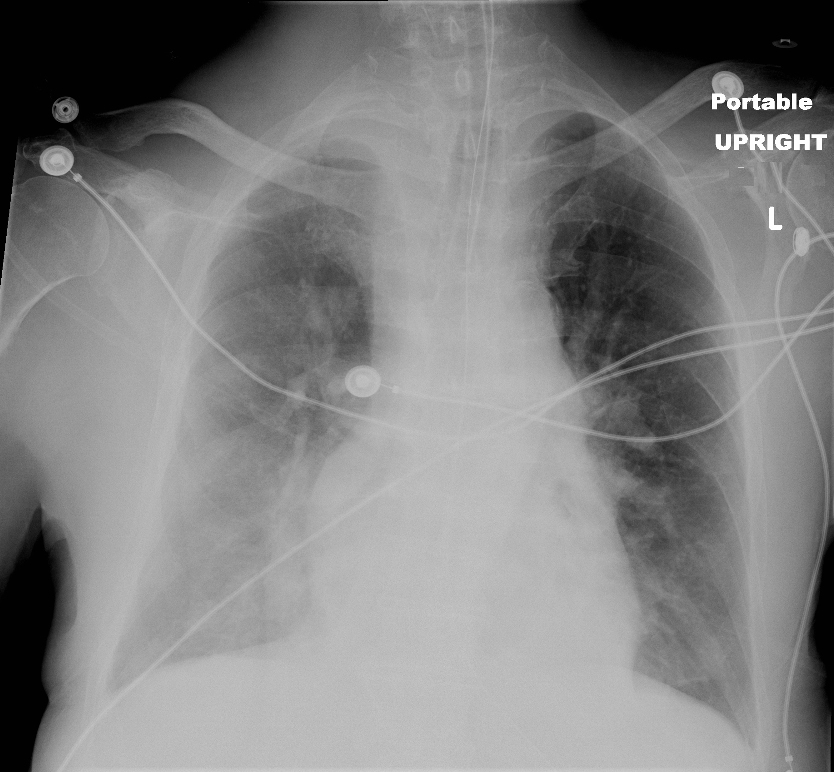
CXR Scimitar Sign and Unilateral Pulmonary Edema
66 year old female presents with dyspnea
Frontal CXR shows unilateral right sided pulmonary edema with cephalization of the left upper lung vessels, with evidence of left atrial enlargement, and enlarged main pulmonary artery suggesting pulmonary hypertension secondary to longstanding left to right shunt. In addition there is a curvilinear structure along the right heart border that is shaped like a sword or a scimitar and the right lung appears smaller than the left. These findings are consistent with scimitar sign and scimitar syndrome
The large shunt results in in increased flow to the right lung and therefore accounts for the unilateral edema. The cephalization on the left lung suggests heart failure
Courtesy Ashley Davidoff MD The CommonVein.net 127H 82595.8
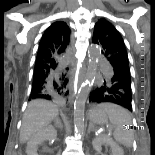
Scimitar Sign and Pulmonary Hypertension
66 year old female presents with dyspnea
Coronal CT shows a curvilinear sword shaped structure in the right lower lung field that represents the right lower pulmonary vein that is draining anomalously into the inferior vena cava.
The pulmonary arteries are enlarged reflecting pulmonary hypertension secondary to longstanding left to right shunt
Findings are consistent with partial anomalous pulmonary venous return and the sword like shape of the vein has been assigned the name scimitar syndrome The PAPVR is often associated with hypoplasia of the lung on the affected side.
Courtesy Ashley Davidoff MD The CommonVein.net 127H 82604
66 year old female presents with dyspnea
Coronal CT shows a curvilinear sword shaped structure in the right lower lung field that represents the right lower pulmonary vein (red arrowhead, b) that is draining anomalously into the inferior vena cava(blue arrowhead b).
The pulmonary arteries are enlarged reflecting pulmonary hypertension secondary to longstanding left to right shunt
Findings are consistent with partial anomalous pulmonary venous return and the sword like shape of the vein has been assigned the name scimitar syndrome The PAPVR is often associated with hypoplasia of the lung on the affected side.
Courtesy Ashley Davidoff MD The CommonVein.net 127H 82604cL
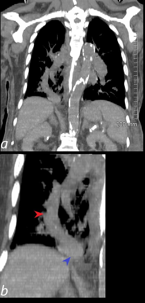
Scimitar Sign on CT
66 year old female presents with dyspnea
Coronal CT shows a curvilinear sword shaped structure in the right lower lung field that represents the right lower pulmonary vein (red arrowhead, b) that is draining anomalously into the inferior vena cava(blue arrowhead b).
The pulmonary arteries are enlarged reflecting pulmonary hypertension secondary to longstanding left to right shunt
Findings are consistent with partial anomalous pulmonary venous return and the sword like shape of the vein has been assigned the name scimitar syndrome The PAPVR is often associated with hypoplasia of the lung on the affected side.
Courtesy Ashley Davidoff MD The CommonVein.net 127H 82604cL
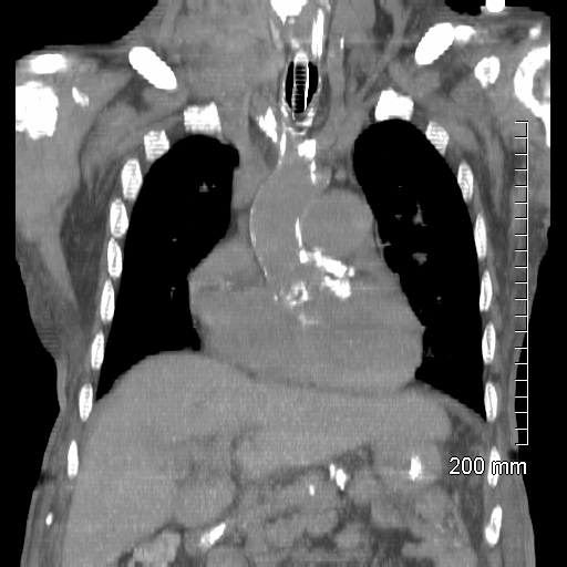
PAPVR and Pulmonary Hypertension
66 year old female presents with dyspnea
Coronal CT shows an enlarged main pulmonary artery measuring 4.2cms reflecting pulmonary hypertension secondary to longstanding left to right shunt. Note the advanced atherosclerotic calcification of the aortic valve and ascending aorta
Courtesy Ashley Davidoff MD The CommonVein.net 127H 82601
66 year old female presents with dyspnea
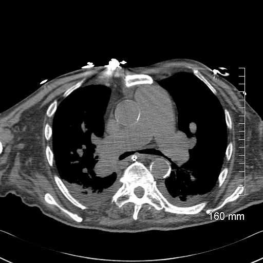
Axial CT shows an enlarged main pulmonary artery measuring 4.2cms reflecting pulmonary hypertension secondary to longstanding left to right shunt. Small Bilateral effusions with compressive atelectasis are present
Courtesy Ashley Davidoff MD The CommonVein.net 127H 82597
66 year old female presents with dyspnea
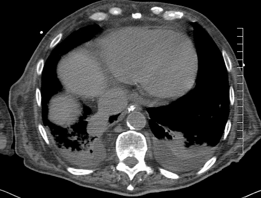
Axial CT shows an enlarged right lower pulmonary vein that is draining anomalously into the inferior vena cava.
Small Bilateral effusions with compressive atelectasis are present
Courtesy Ashley Davidoff MD The CommonVein.net 127H 82600
66 year old female presents with dyspnea
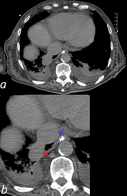
Axial CT shows an enlarged right lower pulmonary vein (red arrowhead, b) that is draining anomalously into the inferior vena cava(blue arrowhead, b).
Small Bilateral effusions with compressive atelectasis are present
Courtesy Ashley Davidoff MD The CommonVein.net 127H 82600cL
Pulmonary Hypertension on MRI Spin Echo Sequence
66 year old female presents with dyspnea
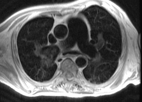
Axial MRI using spin echo(SE) “black blood imaging shows an enlarged main pulmonary artery measuring 4.2cms and enlarged branch pulmonary arteries reflecting pulmonary hypertension secondary to longstanding left to right shunt.
Courtesy Ashley Davidoff MD The CommonVein.net 127H 44626
Pulmonary Hypertension on MRI (SSFP)
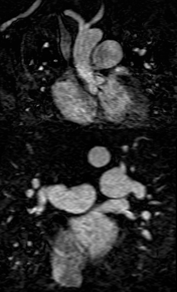
66 year old female presents with dyspnea
Coronal MRI using steady-state free precession (SSFP) shows an enlarged main pulmonary artery measuring 4.2cms and enlarged branch pulmonary arteries reflecting pulmonary hypertension secondary to longstanding left to right shunt.
Courtesy Ashley Davidoff MD The CommonVein.net 127H 44607c
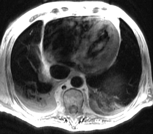
66 year old female presents with dyspnea
Axial MR using “black blood” (SE) sequence shows an enlarged inferior vena cava, which is larger than the aorta. This abnormality is secondary to the known anomalous drainage of the right lower pulmonary vein to the IVC.
Courtesy Ashley Davidoff MD The CommonVein.net 127H 44627