

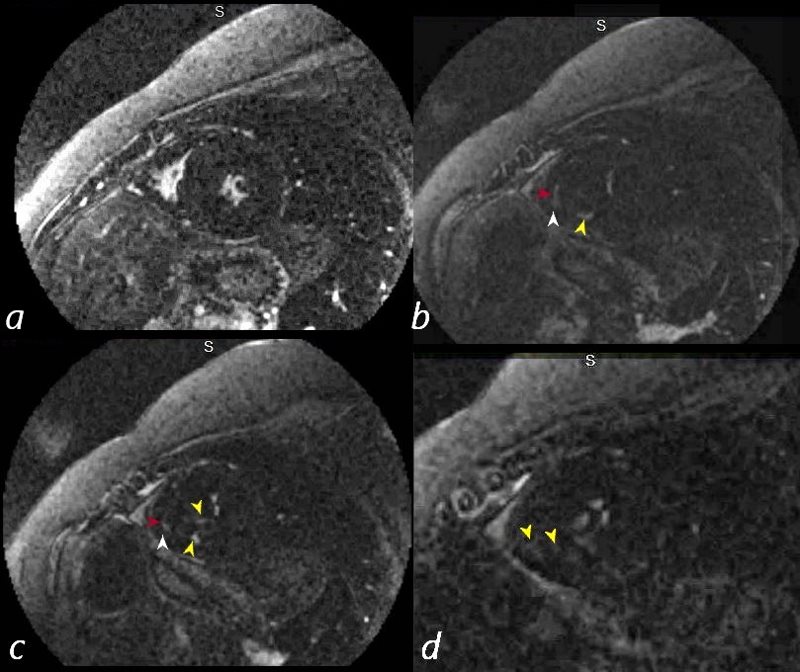
54 year old female with a history of sarcoidosis diagnosed on a groin node biopsy. History of recurrent pericarditis with tamponade s/p pericardial window
MRIs performed 4 months apart reveal vague nodular changes at the hinge points and in the inferolateral portions of the left ventricle. In the first short axis scan the changes were thought to be artifact. However on follow up the finding are persistent and therefore real.
On both scans there is diffuse enhancement of the pericardium.
Courtesy Ashley Davidoff MD
ASYMMETRIC HYPERTROPHIC OBSTRUCTIVE CARDIOMYOPATHY
Ashley Davidoff MD
References and Links
-
-
-
TCV
-
Case Studies
-
-
-