

Ashley Davidoff MD
TheCommonVein.net
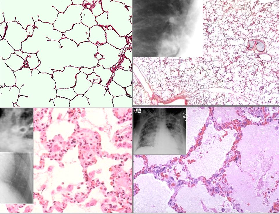
The top left image is the a histological section of normal alveoli and normal wall and interstitium. Heart failure occurs when the left ventricular end diastolic pressure rises. There are 3 basic phases of heart failure. in the first phase (top right) the LVEDP rises above 12 mmHg and on an upright CXR there is equalization of the size of the vessels going to the upper lobes and lower lobes. As the LVEDP goes above about 15-18 mm Hg there is cephalization of the vessels and the upper lobe vessels are larger than the lower lobe vessels.
The second phase of interstitial edema (bottom left) occurs when the intravascular hydrostatic pressure exceeds the intravascular oncotic pressure and this occurs when the LVEDP goes above 25 mm Hg. Fluid accumulates in the alveolar walls and interstitium and the wall becomes thicker with fluid, and the lymphatics and interlobular septa are distended.
The last phase of alveolar edema (lower right) occurs when the pressure exceeds 35 mmHg and the fluid leaks into the alveoli .
Ashley Davidoff MD
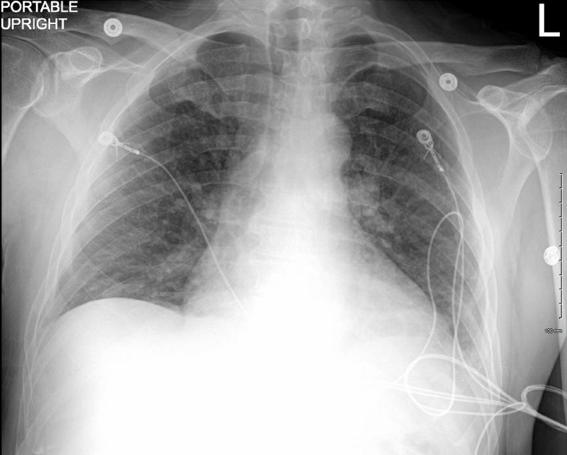
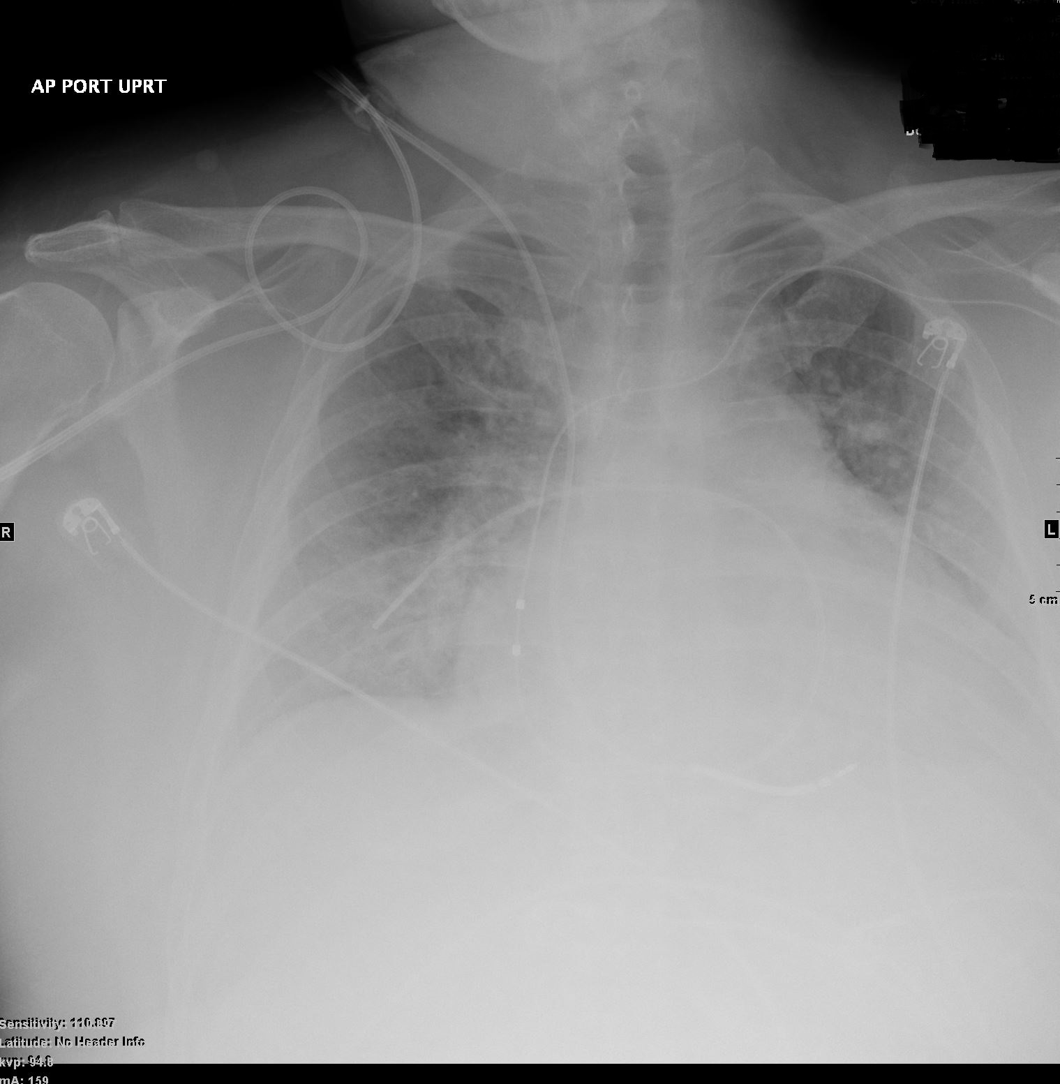
55-year-old male with no prior history became unresponsive during sexual intercourse. Required CPR and defibrillation, and subsequently required epinephrine drip
Echo showed EF 50-55%, and normal cardiac chambers.
CXR showed CHF and LAE
Cardiac Cath showed normal coronaries, PCW 25 mm Hg
MRI showed mildly dilated LV and normal sized left atrium, right atrium and right ventricle. There was LGE in the mid myocardium in the apical anterior wall . Ejection fraction was 47% and myocardial mass was 61g/sqm. End diastolic volume was 160 ccs. Associated findings included bilateral pleural effusions. Finding were most consistent with a myocarditis, or sarcoidosis. Also included in the differential diagnosis was methamphetamine analogue use.
CXR following placement of a pacemaker/defibrillator showed improved CHF
Ashley Davidoff MD
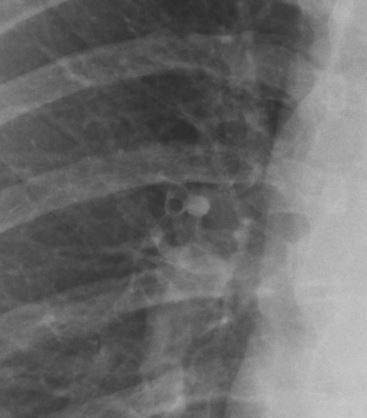
The Normal Segmental Airway Wall Thickness
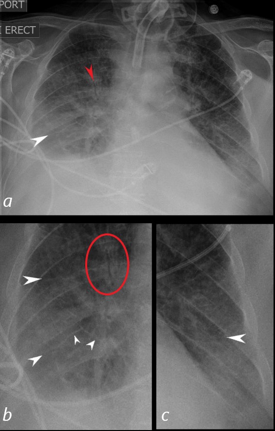
62-year-old male in the ICU with a tracheostomy with acute respiratory distress.
CXR shows acute alveolar edema with an air bronchogram in the right upper lobe (red arrow, a), and red circle, b)with Kerley A lines extending from the periphery to the hila and mediastinum (white arrows)
Ashley Davidoff MD
Peribronchial Cuffing

62-year-old male in the ICU with a tracheostomy and in acute respiratory distress.
CXR shows acute alveolar edema with an air bronchogram in the right upper lobe (red arrow, a), and red circle, b) with peribronchial cuffing around bilateral mid lung bronchi (teal arrows)
Ashley Davidoff MD
Presented with Interstitial edema
After Pacemaker improved

