
Acute Myocardial Infarction
The Common Vein Copyright 2008
Definition
Myocardial infarction is a circulatory disorder of the heart caused by acute coronary occlusion usually by thrombus or by a ruptured plaque that secondarily accumulates platelet thrombi, and results in necrosis and myocardial injury. It is clinically characterized by presence of symptoms of acute myocardial ischemia (ischemic chest pain or chest pain equivalent as dyspnea, diaphoresis, lightheadedness, palpitations).
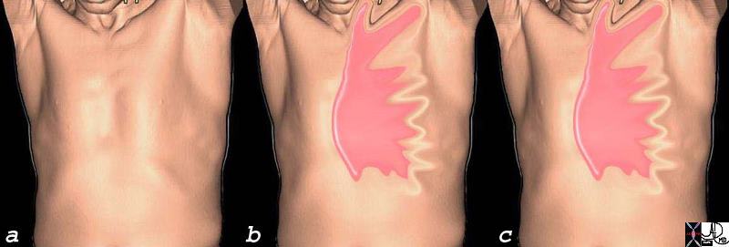
(b) Sudden Severe Pain Pressure Discomfort
c) Unrelieved by TNG and Rest
71197c08c.800 substernal chest pain burning esophagus heart cardiac esophagitis reflux retrosternal chest pain radiating to neck and left arm unrelieved by rest and sublingual nitroglycerine myocardial infarction time hours CTscan 3D Courtesy Ashley Davidofff MD
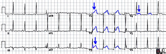
The diagnosis is confirmed by characteristic ECG changes and biochemical markers of myocardial necrosis as reflected by typical rise and fall of cardiac enzymes: troponin I or T, CK-MB and is classified as ST elevation (STEMI)or non ST elevation myocardial infarction (NSTEMI) based on the EKG
The pathological diagnosis of myocardial infarction requires evidence of myocyte cell death (i.e. necrosis of the myocardium) as a consequence of prolonged ischemia.(Luepker)
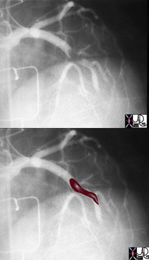
The above image of this cardiac catheterization shows a coned down left anterior oblique (LAO) projection of the LAD in a patient with acute thrombosis of the left anterior descending artery. In the lower image, the thrombus in the artery is overlaid in maroon.
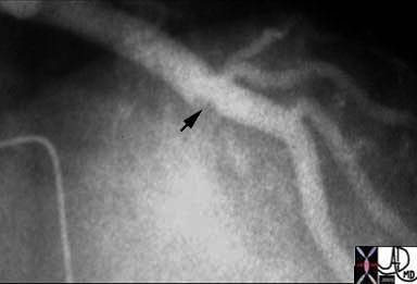
Following thrombolysis, minimal irregularity (black arrow) of the endothelium is seen. This may be residual thrombus or the remnants of a plaque rupture.
Images Courtesy of: Ashley Davidoff, M.D.
Treatment depends on the type of infarction (STEMI vs NSTEMI, but attempts are usually made to lyse thrombus and open the vessels by angioplasty or stenting in a timely manner in order to salvage myocardium at risk.
Ashley Davidoff MD
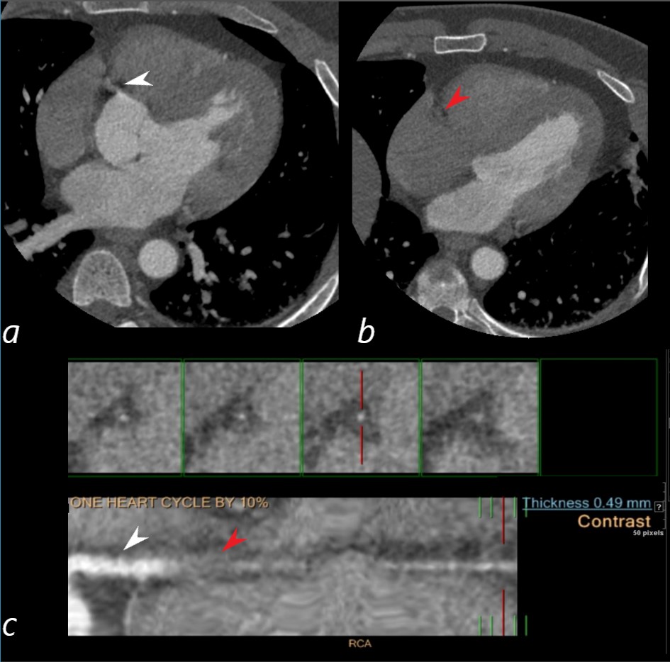
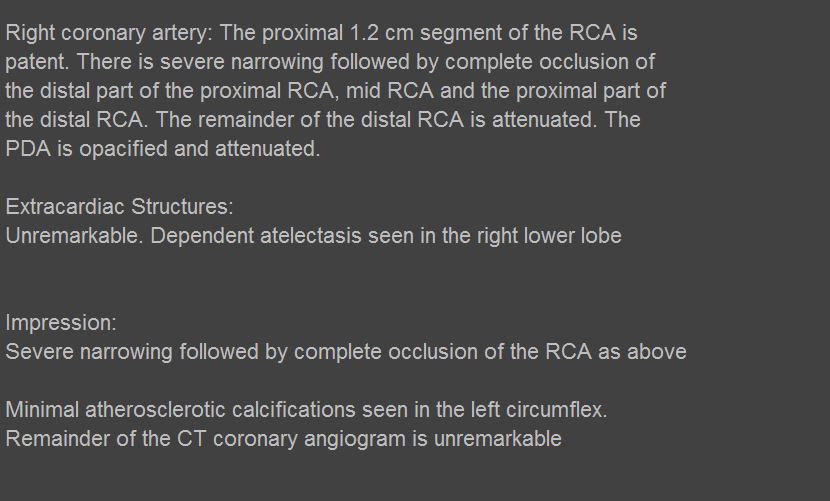
009-30-m-heart-RCA-occlusion c01
Ashley Davidoff MD
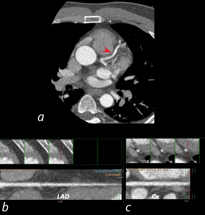
018-30m-heart-LAD Cx patent
Unusual Cases
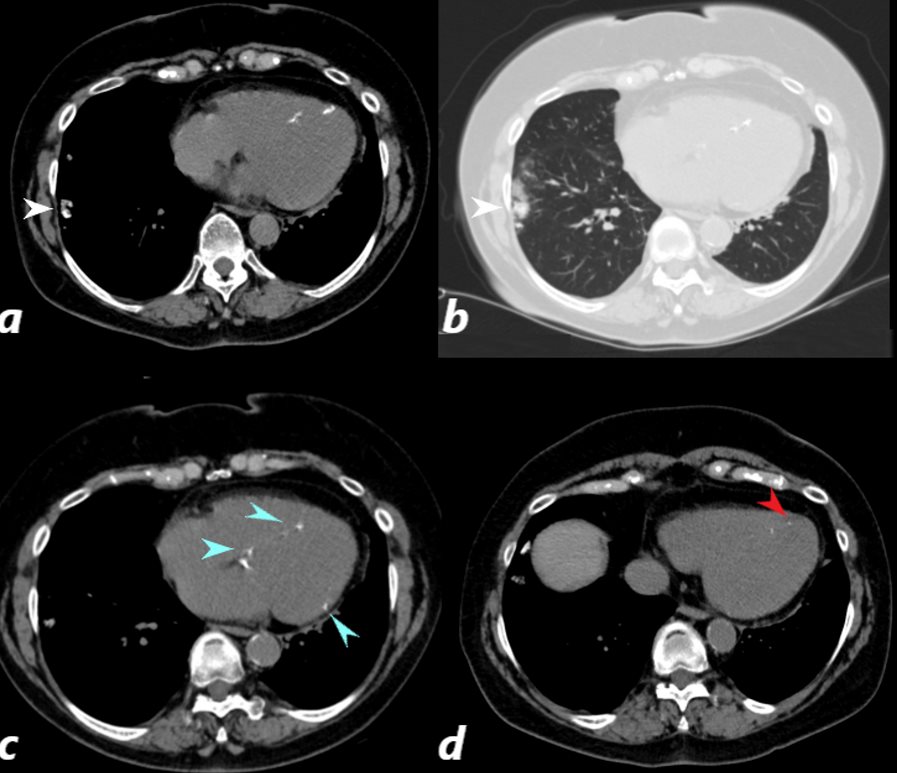
CT scan of a 67 year old female with anca vasculitis shows regions of dystrophic calcification in the lateral aspect of the right lower lobe (white arrow, a and b) )with focal nodular parenchymal consolidation, that likely reflects a site of prior small vessel infarct. Dystrophic calcification in the LV myocardium (blue arrows c) and a suggestion of fatty dysplasia in the left ventricular apex red arrow d) suggest changes from small vessel infarct. Ashley Davidoff MD
References
- Luepker RV, Apple Fs et al: Case definitions for acute coronary disease in epidemiology and clinical research studies. Circulation 108:2543, 2003