
Chambers that are Border Forming on the PA Examination
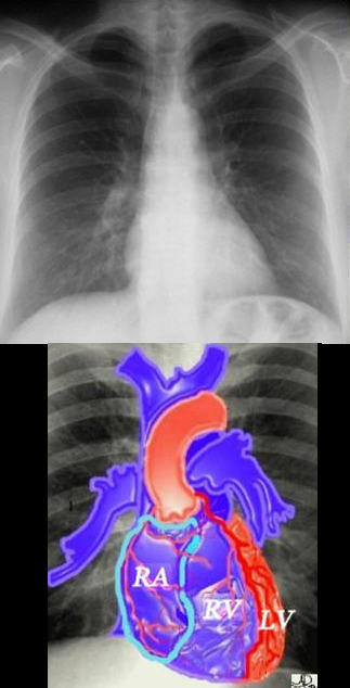
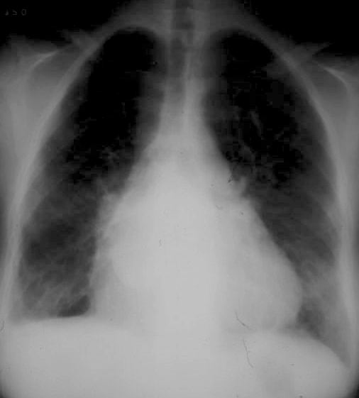
If we were to “crack open” the chest of the chest X-ray, the structures that would dominate this bloody, black and white scene, would be the right sided chambers. The right ventricle (RV) would be the dominant anterior chamber, and would form the dominant interface with the diaphragm. The right atrium (RA) would form the border with the right lung. The RA would of course be slightly posterior to the RV. The left border would be formed by the left ventricle. Most the left ventricle is hidden posteriorly in this view. The left anterior descending artery would be visible from this anterior view. It marks the position of the interventricular septum.
Ashley Davidoff MD
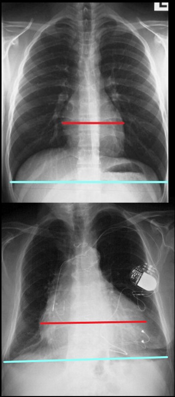
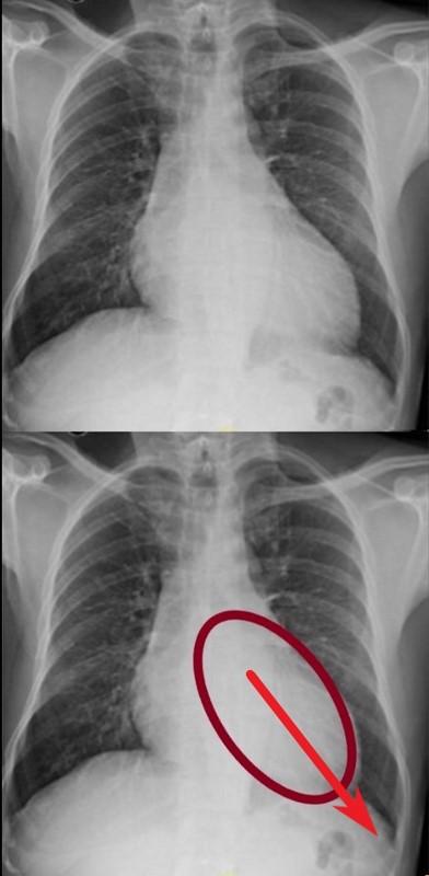
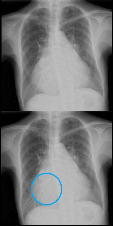
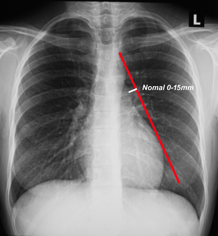
The maximum transverse length of the heart is expressed as a percentage of the maximum length of the internal diameter of the chest. When this ratio – the cardiothoracic ratio (c t r) is greater than 50% cardiomegaly is present. The top image is normal and the bottom reflects cardiomegaly
Ashley Davidoff MD
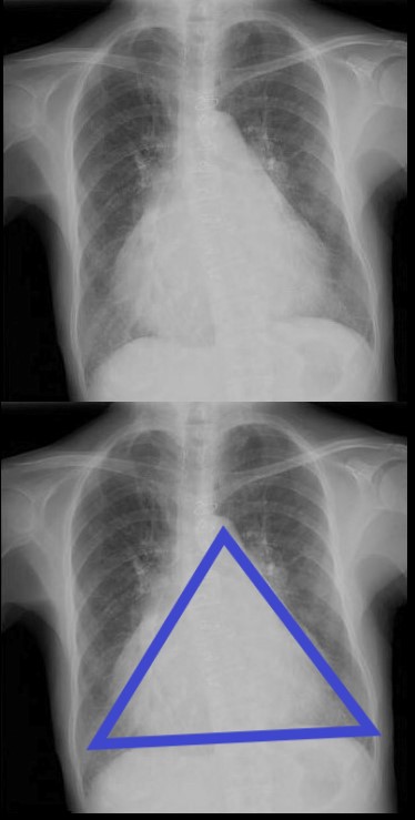
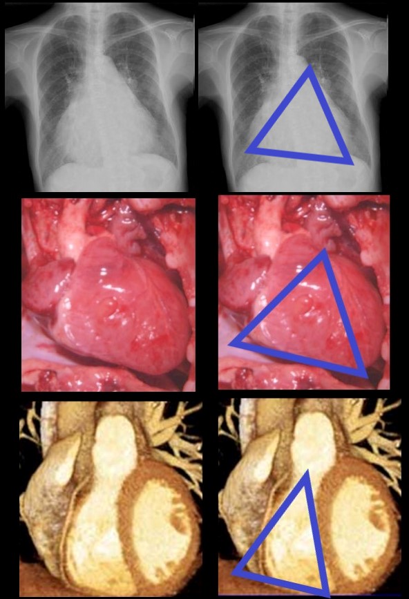
There are Two Basic Common Shapes of Cardiomegaly
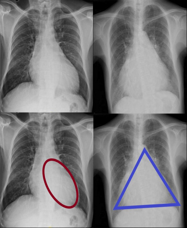
CARDIOMEGALY – TWO BASIC TYPES -OVOID and TRIANGULAR
The ovoid form which suggests left ventricular dominance and triangular form which suggests right ventricular dominance.
Ashley Davidoff MD
The Ovoid Form of Cardiomegaly – Consider LVE
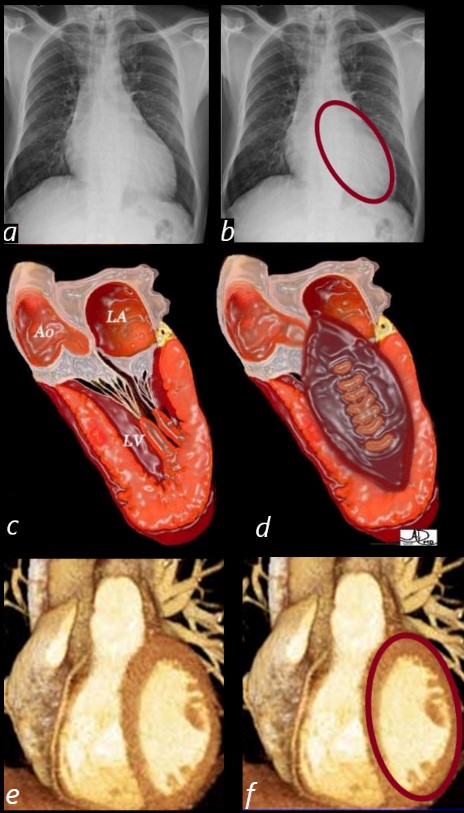
SHAPE OF THE LEFT VENTRICLE
The enlarged LV (a,b) is shaped like an oval and it is likened to a rugby ball or an American football placed on the field at kick off time. LVE on CXR is mostly assessed by an increased cardiothoracic ratio as well as the accentuation of the ovoid shape. (lower images c, d,e, f)
Ashley Davidoff MD
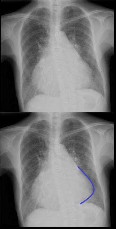
Vector of the Enlarging LV – Rotate Down and to the Left
 VECTOR FOR LV ENLARGEMENT
DOWN AND OUT
The left ventricle (LV) enlarges in a posterior, downward and lateral direction resulting in the characteristic changes of LVE on CXR
Ashley Davidoff MD
Clinical Exam – Focal LV Thrust
The left ventricle (LV) enlarges in a downward and lateral direction resulting in the apical impulse displacement and increase forcefulness of the apical tap.
Ashley Davidoff MD
62 year old female with acute chest pain atrial fibrillation, hypotension admitted to ICU. Clinical evaluation was considered to be non-ischemic cardiomyopathy with EF by echo of about 20%. She was hypotensive and, in the ICU, and CXR showed acute CHF with cardiomegaly. The TEE was more in keeping with segmental dyssynergy, Cardiac cath showed occluded RCA bot good collateralization from the LAD. MRI showed subendocardial LGE in the inferior and inferolateral portions of the LV consistent with a prior infarction and EF of 20%
Ashley Davidoff MD
The Triangular Form of Cardiomegaly
Consider RVE or any disease from the left side that may cause RVE eg mitral stenosis
TRIANGULAR SHAPED HEART – SUGGESTING RIGHT VENTRICULAR DOMINANCE. MITRAL STENOSIS PULMONARY HYPERTENSION
71 year old Asian female with rheumatic heart disease dominated by calcific mitral stenosis mild MR, moderate tricuspid regurgitation and secondary pulmonary hypertension.
Ashley Davidoff MD
Note the Basic Shape of the RV is Triangular in almost all Views
The enlarged LV (a,b) is shaped like an oval and it is likened to a rugby ball or an American football placed on the field at kick off time. LVE on CXR is mostly assessed by an increased cardiothoracic ratio as well as the accentuation of the ovoid shap. (lower images c, d,e, f)
Ashley Davidoff MD
Vector of the Enlarging RV – Rotate Laterally to the Right
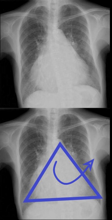
ANTERIOR LEFTWARD
The right ventricle (RV) enlarges with a clockwise rotation resulting in an upward turning of the apex and enlargement in a anterior and leftward lateral direction.
Ashley Davidoff MD
As the right ventricle (RV) enlarges with a clockwise rotation the apex of the small ventricle succumbs to the larger silhouette of the RV which pints upward and to the left and has been called the “proud breast” appearance.
Ashley Davidoff MD

Ashley Davidoff MD
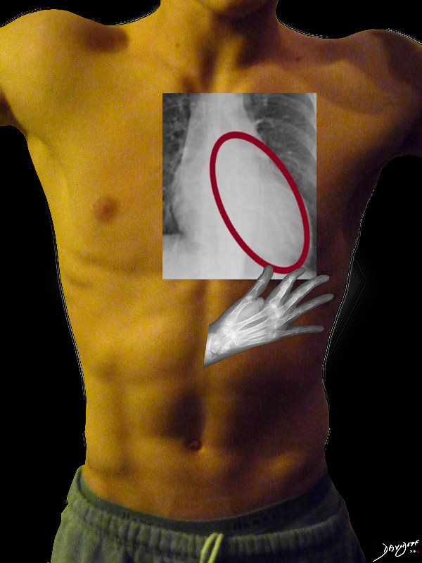
Clinical Exam of the RV – Diffuse Anterior Parasternal Heave
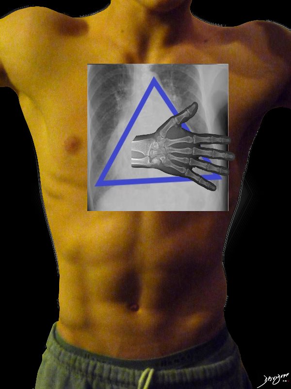
The right ventricle (RV) enlarges in a anterior, upward and lateral direction resulting in the broad based parasternal pulsation which on clinical examination is identified as a parasternal heave identified with the base of the extended hand
Ashley Davidoff MD
Triangular shaped heart with RVE LAE
Ashley Davidoff MD
Frontal x-ray with triangular shaped heart due to pulmonary hypertension with enlarged MPA and enlarged descending RPA .
Ashley Davidoff MD
What about the Right Atrium (RA) and Right Heart Border?
The RA does not make much a statement on the frontal CXR unless very large
If we were to “crack open” the chest of the chest X-ray, the structures that would dominate this bloody, black and white scene, would be the right sided chambers. The right ventricle (RV) would be the dominant anterior chamber, and would form the dominant interface with the diaphragm. The right atrium (RA) would form the border with the right lung. The RA would of course be slightly posterior to the RV. The left border would be formed by the left ventricle. Most the left ventricle is hidden posteriorly in this view. The left anterior descending artery would be visible from this anterior view. It marks the position of the interventricular septum.
Ashley Davidoff MD
The Enlarged Right Atrium
The right atrium is the most difficult chamber to assess unless it is very large in which case it will manifest on the frontal CXR with a very large right paravertebral border.
-
- enlarged, globular heart
- narrow pedicle
- gross enlargement of the right atrial shadow, i.e. increased convexity in the lower half of the right cardiac border
- right atrial convexity is more than 50% of the cardiovascular height
- right atrial margin is more than 5.5 cm from the midline
The right atrium is the most difficult chamber to assess unless it is very large in which case it will present on the frontal CXR with a very large right paravertebral border. This is a 71 year old female person with rheumatic heart disease with pulmonary hypertension and tricuspid regurgitation hence resulting in a large right atrium (RAE)
Ashley Davidoff MD
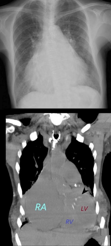
The right atrium is the most difficult chamber to assess unless it is very large in which case it will present on the frontal CXR with a very large right paravertebral border. The frontal CXR and coronal CT through the RA is from a 71 year old female with rheumatic heart disease with pulmonary hypertension and tricuspid regurgitation resulting in a giant right atrium (RAE). The RA accounts for the large bulge of the right border of the cardiac silhouette. The black arrowhead in the loer image points to the calcified mitral valve.
Ashley Davidoff MD
The Left Atrium
3 signs
-
- Carinal Angle
- Double Density
- Filling in of the Left Atrial Appendage Bay
The Carina and the Carinal Angle
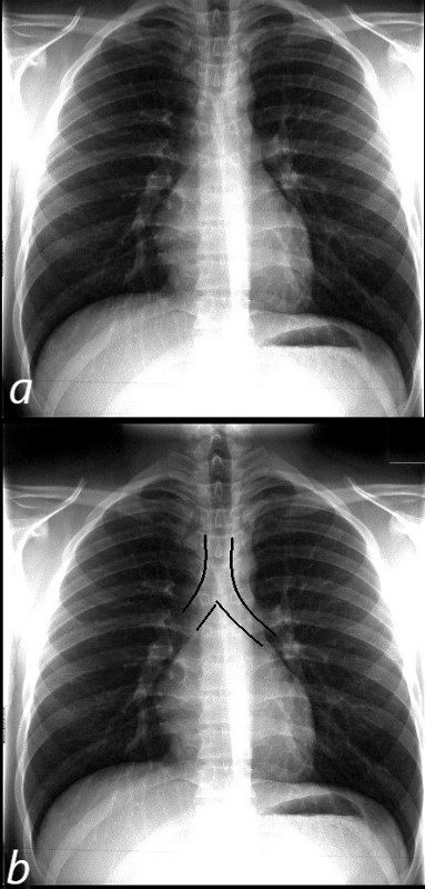 NORMAL FRONTAL CXR NORMAL ASYMMETRIC BRANCHING OF MAINSTEM BRONCHI
The normal CXR shows the characteristic asymmetric branching of the main stem bronchi. The right is short and stout and slightly more vertical while the left is long and thin and slightly more obtuse.
The normal carinal angle is between 40-80 degrees.
Ashley Davidoff MD
 ASYMMETRIC BRANCHING PATTERN – RIGHT SHORT AND STOUT AND THE LEFT LONG AND THIN
CARINAL ANGLE – 40-80 degrees
Ashley Davidoff MD
The Abnormal Carinal Angle

A dancer demonstrates a normal carinal angle (upper image) and as she continues to extend her left leg, (lower images) the angle becomes greater than 80 degrees and in terms of the carinal angle becomes abnormal.
Ashley Davidoff MD
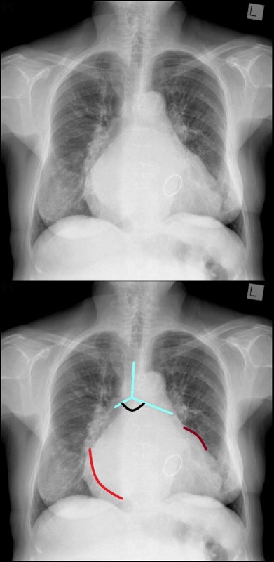
MITRAL STENOSIS WITH ENLARGED LEFT ATRIUM – WIDENED CARINAL ANGLE DOUBLE DENSITY ENLARGED LEFT ATRIAL APPENDAGE
The frontal CXR demonstrates findings consistent with mitral stenosis including a widened carinal angle (teal blue and black arc), a double density (red arc) and an enlarged left atrial appendage (maroon arc).
The overall shape of the heart is triangular suggesting right ventricular enlargement. A mitral valve prosthesis is in position
Courtesy of Radiopaedia
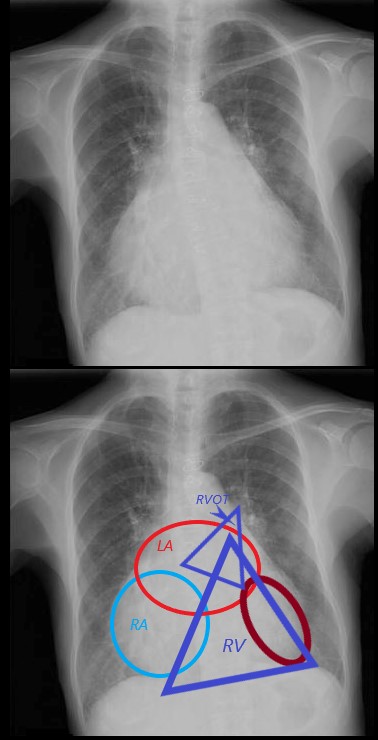
PARTS OF THE ENLARGED HEART ON FRONTAL CXR in MITRAL STENOSIS PULMONARY HYPERTENSION AND COR BOVINUM
71 year old Asian female with rheumatic heart disease dominated by calcific mitral stenosis mild MR, moderate tricuspid regurgitation and secondary pulmonary hypertension.
The frontal view shows an enlarged RA (light blue) characterized by a prominent right heart border. The right ventricle is enlarged as noted by the triangular shape of the cardiac silhouette, an the upturned (“proud breast”) appearance of the left heart border, both reminiscent of RVE. The left atrium is also significantly enlarged characterised by the widened carina and the straight heart border caused by a combination of atrial appendage enlargement and pulmonary hypertension. The LV (maroon) is normal in size
Ashley Davidoff MD
|
The Shapes of the Heart in Health and Disease |
|
From top left ti right and across the rows they are: The normal heart , the “football” of LV enlargement the “triangle” or “proud breast” of RV enlargement, “snowman” of total anomalous pulmonary venous return, big PA mogul of pulmonary hypertension, “egg on its side” of D transposition of the great vessels, “boot shaped” heart seen in both pulmonary atresia and Tetralogy of Fallot, the long smooth combined Ao and PA mogul that has a differential diagnosis of L transposition, absence of the pericardium, and juxtaposition of the atrial appendages, the box shaped large heart of Ebstein’s anomaly, dextrocardia , and the water bottle” heart of a large pericardial effusion. 07197 Images are a combination of images from a personal collection and borrowed from the internet for educational purposes only. Some of the sources are unknown and are used for educational purposes alone 86774b02 |
Normal Pulmonary Artery
When a line is drawn from the aortic knob to the left edge of the heart, (red line) the pulmonary artery should lie medial to that line (ie along the line drawn to 1.5cms medial to the line)
Ashley Davidoff MD TheCommonVein.net
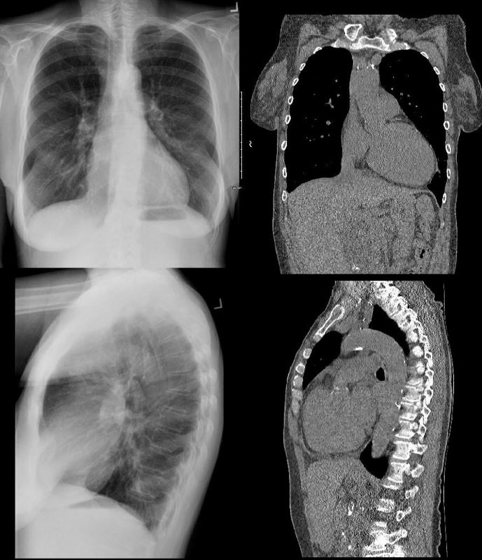
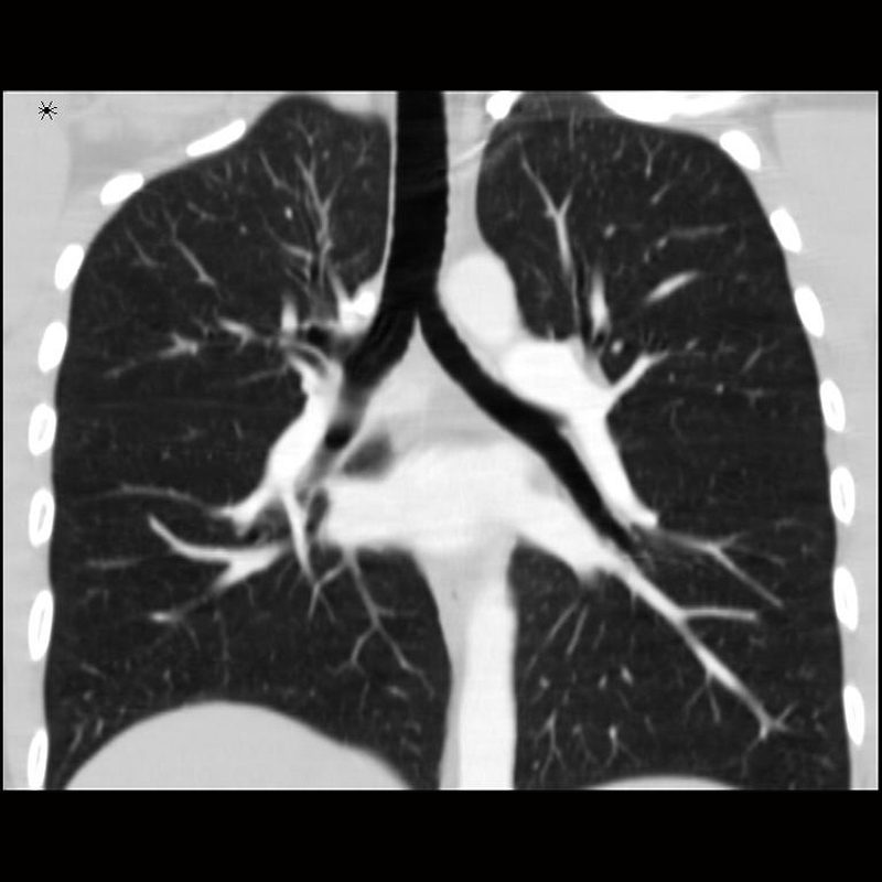
Pulmonary Hypertension
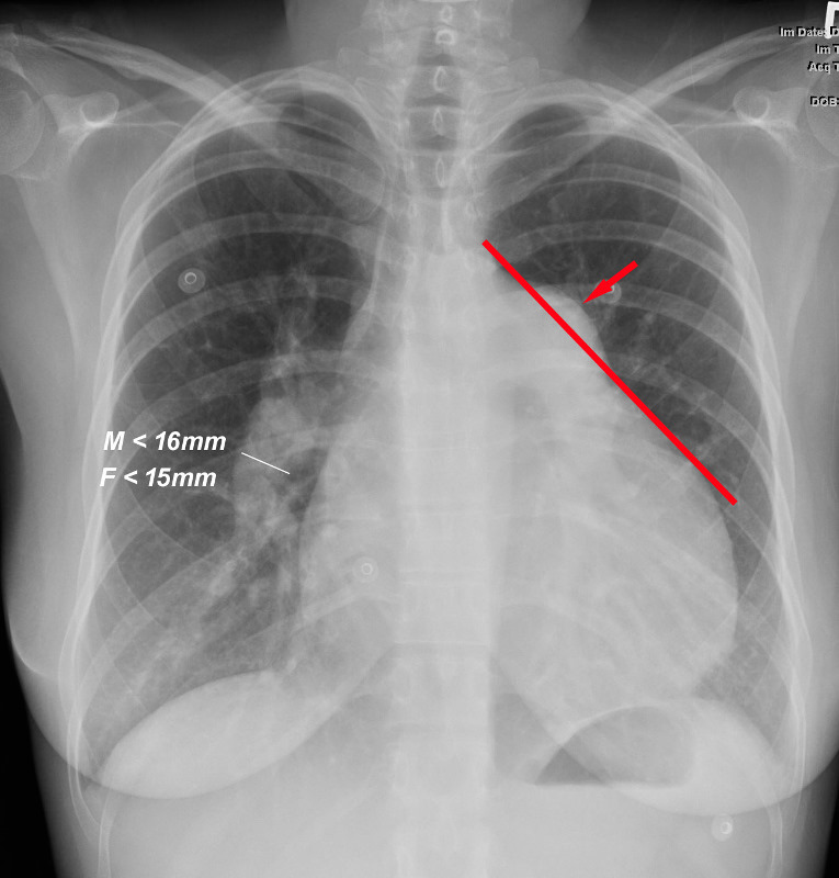
When a line is drawn from the aortic knob to the left edge of the heart, (red line) the pulmonary artery lies lateral to that line indicating an enlarged pulmonary artery most commonly caused by hypertension . In this instance the size of the descending right pulmonary artery is greater than 15 mms confirming the presence of pulmonary hypertension
Ashley Davidoff MD TheCommonVein.net