75-year-old female with a history of non-ischemic restrictive cardiomyopathy, EF 30%, cardiogenic shock, atrial fibrillation, status post myocardial biopsies, hypertension hyperlipidemia, rheumatoid arthritis hypothyroidism, mitral valve prolapse, past history of Takotsubo syndrome
Right heart cath showed RA pressure of 13, RV of 43/8, PA 46/26 PCWP of 24, CI of 1.3
CXR 1 year ago with cardiomegaly and mild to moderate CHF
CXR 1-year prior shows cardiomegaly, with left atrial enlargement, cephalization and early interstitial edema consistent with moderate CHF Ashley Davidoff MD 116792a
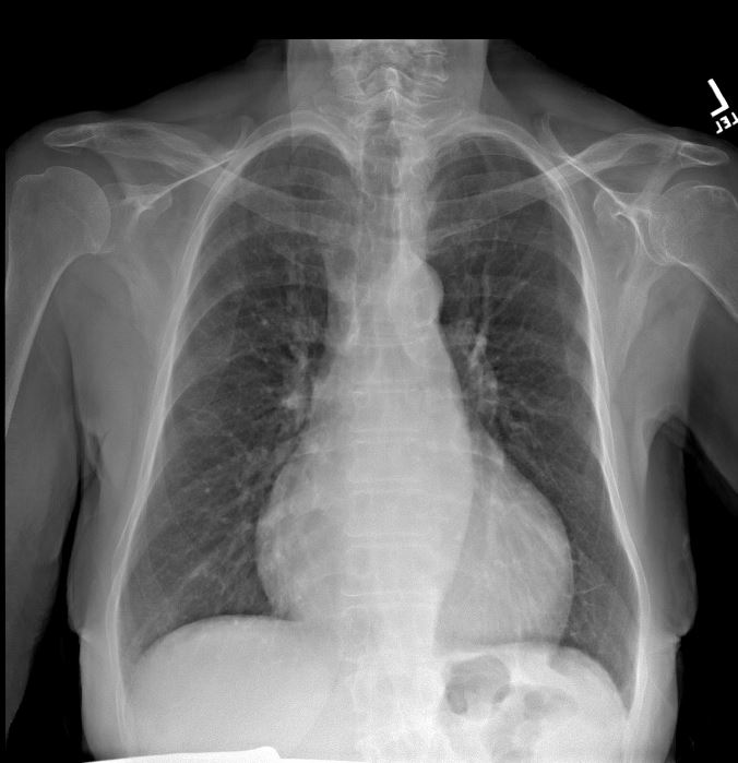
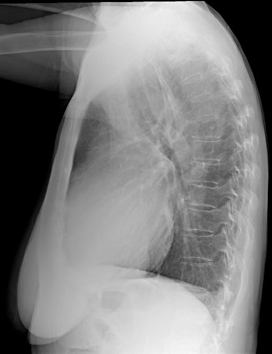
CXR 3 months prior with cardiomegaly and mild CHF
CXR in 2 views 3 months earlier shows cardiomegaly with left atrial enlargement, LV enlargement (lateral exam) cephalization without interstitial edema consistent with mild CHF Ashley Davidoff MD 116792b01 and 116792b01
CXR in 2 views 3 months earlier shows cardiomegaly with left atrial enlargement, LV enlargement (lateral exam) cephalization without interstitial edema consistent with mild CHF Ashley Davidoff MD 116792b01 and 116792b01
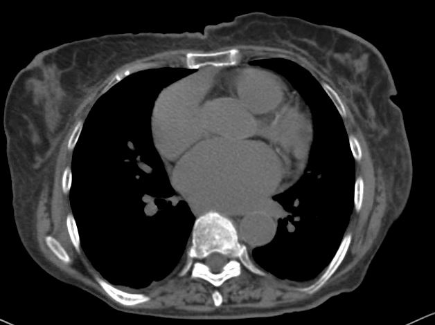
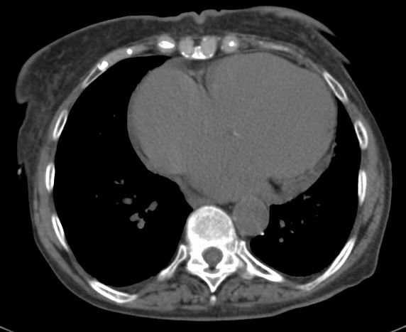
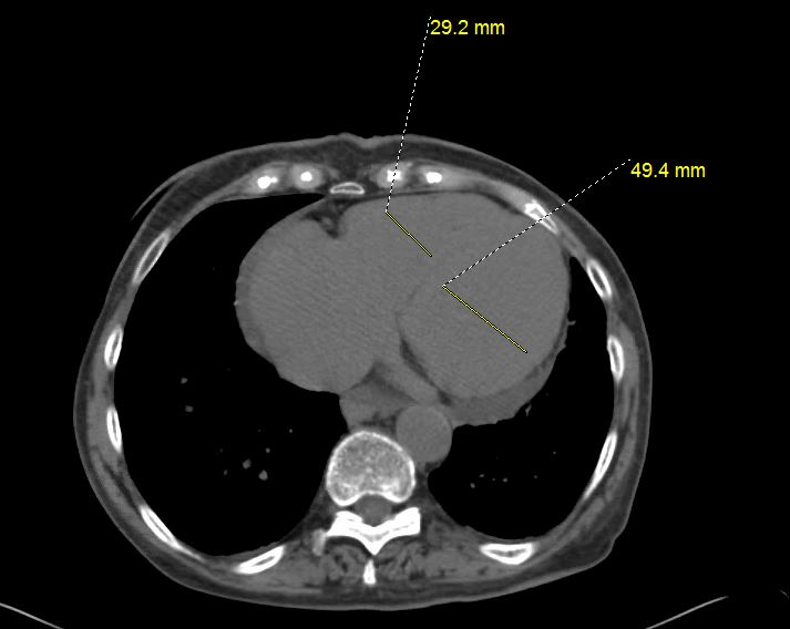
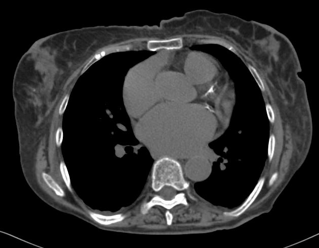
CT scan 1 month prior showing bi atrial enlargement LVE, early pulmonary hypertension and small pericardial effusion
Axial CT scan through the left atrium shows enlargement of the left atrium Ashley Davidoff MD 116792b04Axial CT scan through the level of the right atrium shows enlargement of the right atrium and a small pericardial effusion, low density blood consistent with anemia Ashley Davidoff MD 116792b05Axial CT scan through the level of the LV shows enlargement of the left ventricle, a small pericardial effusion, low density blood consistent with anemia, and a suggestion of fat in the ventricular septum most often associated with a prior myocardial infarction Ashley Davidoff MD 116792b06Axial CT scan through the level of the LV suggests enlargement of the left ventricle (5cms is upper limits normal) , normal sized RV, a small pericardial effusion, and low density blood consistent with anemia. Ashley Davidoff MD 116792b07Axial CT scan through the level of the coronary arteries shows mild to moderate atherosclerotic calcification of the proximal LAD and circumflex as well as LA enlargement Ashley Davidoff MD 116792b10
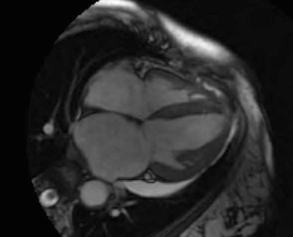
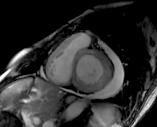
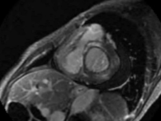
T2 Axial MRI through the level of the LV shows edema of the apex of the LV and a small pericardial effusion. Ashley Davidoff MD 116793
Mild Mitral Regurgitation
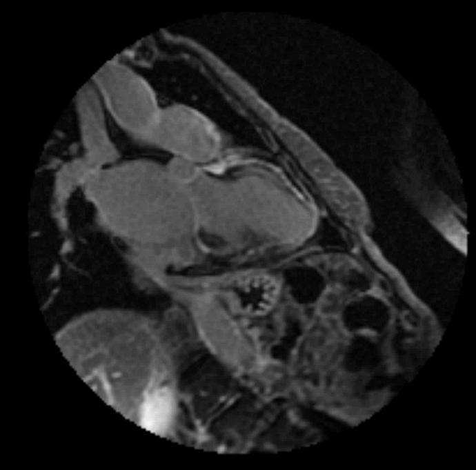
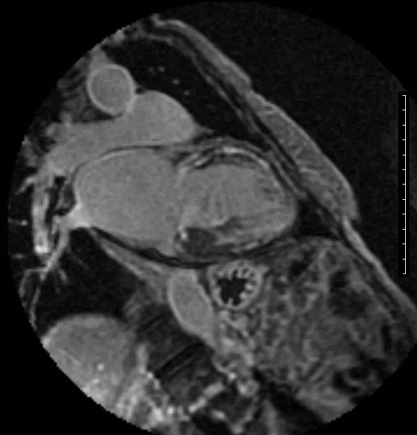
T2 Axial MRI through the level of the LV shows edema of the apex of the LV and a small pericardial effusion. Ashley Davidoff MD 1167934 Chamber MRI through the level of the LV and LA shows biatrial enlargement as well as RV thickening Ashley Davidoff MD 116793b02 4CMRI in the short axis through the level of the LV and LA shows RV thickening and a pericardial effusion Ashley Davidoff MD 116793b03 short axis
MRI, LGE series in the short axis through the level of the LV and RV shows mid ventricular LGE in the left ventricular septum extending to the anterior and anterolateral wall. Ashley Davidoff MD 116797bMRI, LGE series in the short axis through the level of the LV and RV shows mid ventricular LGE in the left in the anterior wall of the LV as well as the free wall of the thickened RV. There are patchy changes in the inferior wall in mid ventricular location Ashley Davidoff MD 116797b01MRI, LGE series in the short axis through the level of the LV and RV shows mid ventricular LGE in the anterior wall of the LV Ashley Davidoff MD 116796MRI, LGE series in the long axis through the level of the LV shows mid ventricular LGE in the left in the anterior wall of the LV as well as inferior wall of the thickened LV. RV free wall is also involved with LGE noted in the free wall Ashley Davidoff MD 116796MRI, LGE series in the long axis through the level of the LV shows mid ventricular LGE in the left in the anterior wall of the LV as well as inferior wall of the thickened LV. RV free wall is also involved with LGE noted Ashley Davidoff MD 116797
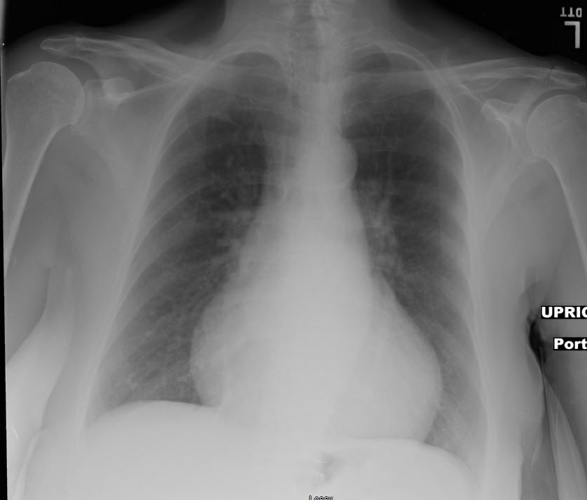
1 Month Later – Moderate CHF
Portable upright AP chest 1 month later shows cardiomegaly with left atrial enlargement and LVE with cephalization and early interstitial edema consistent with moderate CHF Ashley Davidoff MD 116799b01
2 Months Later Mild CHF
Portable upright AP chest 1 month later shows persistent unchanged cardiomegaly with left atrial enlargement and LVE with cephalization and early interstitial edema consistent with moderate CHF Ashley Davidoff MD 116799b01