
Notes
As we scan from superior to inferior
-
NOTES OF THE HEART ON CT
As we start from top to bottom we scan through the PA, aorta, then LA RA, RV and LV
Ashley Davidoff MD
Scales
Going superior to inferior and clockwise
1st stop Aorta LA
-
-
-
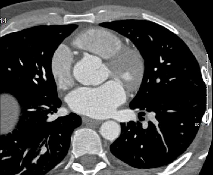
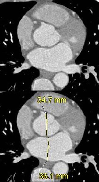
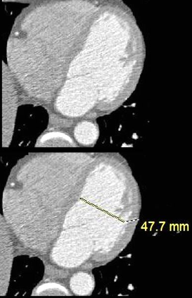
FIRST STOP
Max AP dimension of the LA in the region of the aorta. At this point the LA looks rectangular
Ashley Davidoff MD
-
-
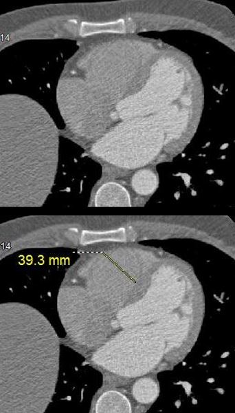
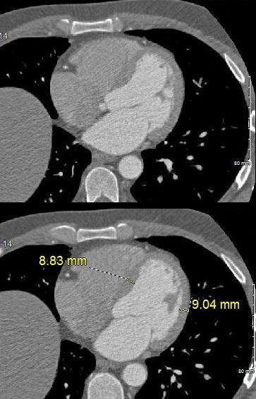
2nd Stop
4 chamber
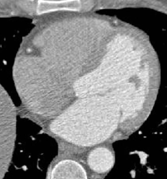
Axial images through the 4 chambers at the level of the A-V valves during diastole (mitral valve open) enables an approximate volume evaluation of the chambers. The atria are approximately the same volumes, and are about 1/3 the volume of the ventricles. The right ventricle (RV) is about 2/3 the volume of the left ventricle (LV)
Ashley Davidoff MD
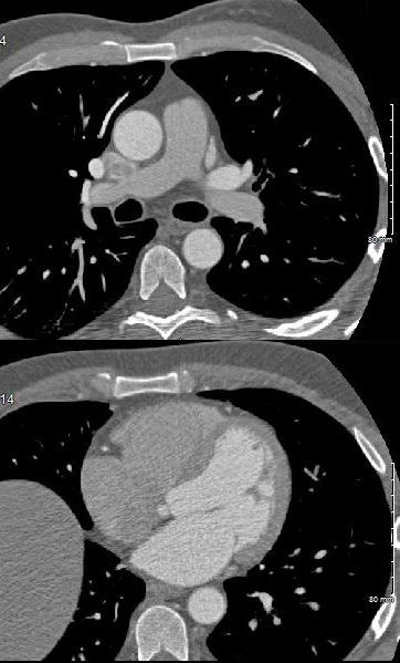
3rd Stop MPA and Bifurcation
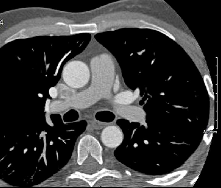
MPA – Aorta
Ashley Davidoff MD
What to do at each stop
Stop 1
Stop 2
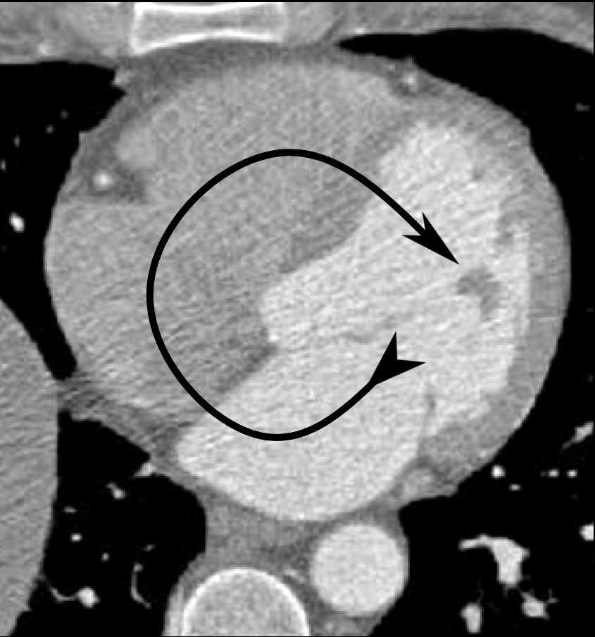
Clockwise Rotation
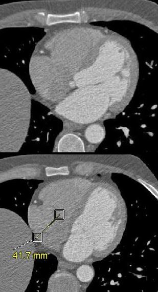
Axial images through the body of the right atrium (RA) at the level of the tricuspid valve shows a linear dimension of 4.2cms (normal up to about 5cms)
Ashley Davidoff MD
Get a Sense of Relative Volumes
3rd Stop PA and Aorta

Practice Practice Practice
- Music
- LAE
- RAE
- RVE
- RV dilated
- RVH
- LVE
- Dilated LV
- LVH
Next Neha Khemani, Size of the Structures of the CV System