

54 year old female with a history of sarcoidosis diagnosed on a groin node biopsy. History of recurrent pericarditis with tamponade s/p pericardial window
MRIs performed 4 months apart reveal vague nodular changes at the hinge points and in the inferolateral portions of the left ventricle. In the first short axis scan the changes were thought to be artifact. However on follow up the finding are persistent and therefore real.
On both scans there is diffuse enhancement of the pericardium.
Courtesy Ashley Davidoff MD
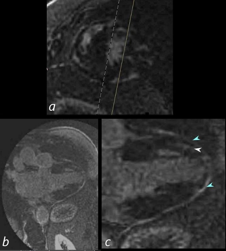
69-year-old male presented with history of cardiomyopathy and atrial fibrillation
In this series, the long axis 2 chambered view (c is magnified view of b) reveals intense continuous pericardial disease (teal blue arrowheads) and mid myocardial linear-nodular LGE (white arrowhead)
Ashley Davidoff MD