
Copyright 2020
- How to diagnose constrictive pericarditis on CT
- Questions
-
- What are the signs of constrictive pericarditis on CT
- pericardial thickening
- diffuse or localized(2mm = normal – 3mm equivocal >3mm abnormal)
- symptoms related to decreased cardiac output in DOE or at rest fatigability a or at rest, or both. impaired diastolic filling of the right ventricle
- signs of fluid overload (ranging from peripheral edema to anasarca),
- ie signs of right heart failure including
- dilatation of the
- RA
- IVC and hepatic veins
- Coronary Sinus
- Azygos vein
- Hepatomegaly
- dilatation of the
- pericardial thickening
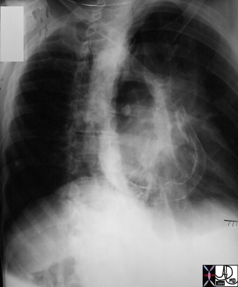
- What is the role of the CXR and LA in Dx
- increase in left atrial because it cannot fill therefore
- increase in pulmonary venous pressure
- Usually normal size LA
- However since LA hmay be only partly covered by pericardium, it may in fact enlarge
- What are the signs of constrictive pericarditis on CT
-
- Questions
Atlas
|
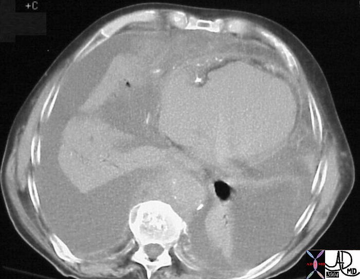
Complications of Hemopericardium |
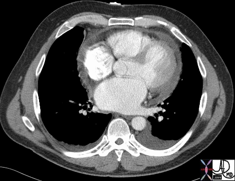
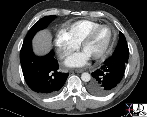
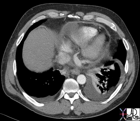
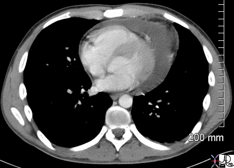
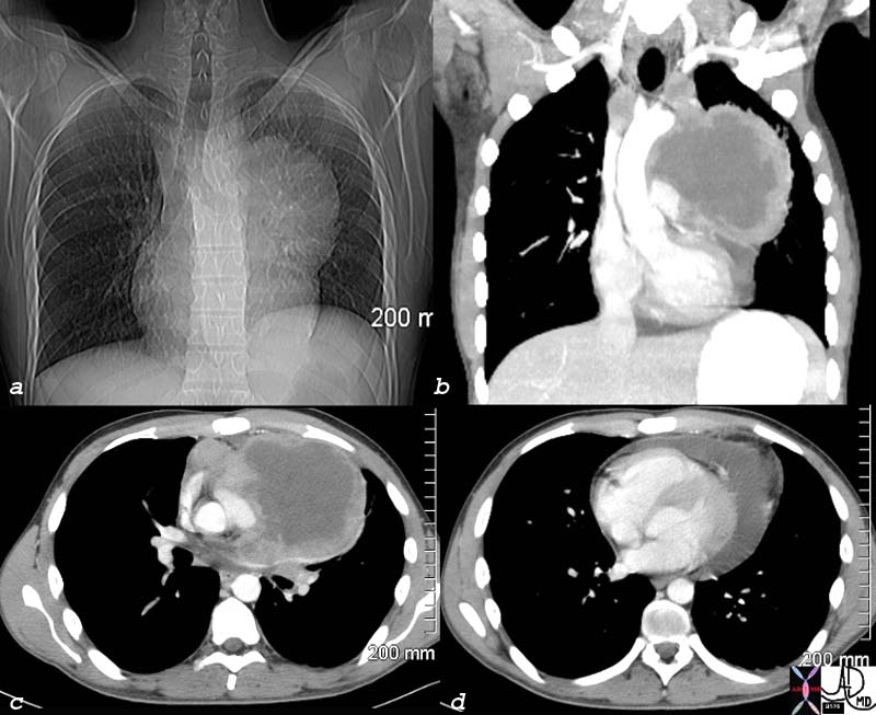
| 43695 51M s/p ablation therapy for atrial fibrillation presents 3 weeks later with SOB pericardium fx small complex pericardial effusion fx thickened complex dx constrictive pericarditis caused by hemopericardium imaging radiology CTscan Courtesy Ashley Davidoff MD |
|
TB pericarditis Calcification of the Visceral and Parietal Pericardium |
| A double layered thin pericardial membranes are turned into a single thick, rigid fibrous capsule, which restricts diastolic filling of the heart and leads to right sided congestive failure. This is usually due to the healing of tuberculosis or purulent bacterial pericarditis.
Courtesy Lloyd Hawes MD 36620 copyright 2009 |
|
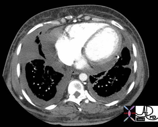
Complications of Breast Carcinoma |
| 39607 Courtesy Ashley Davidoff MD code breast hx SOB fx interstitial thickening fx prominent interlobular septa code heart pericardium fx effusion dx breast carcinoma question lymphangitis question pericardial tamponade CTscan |
Peritoneal Amyloidosis |
| 15528 heart + pericardium + fx calcification + amyloid + imaging radiology CTscan infiltrative amyloidosis |
|
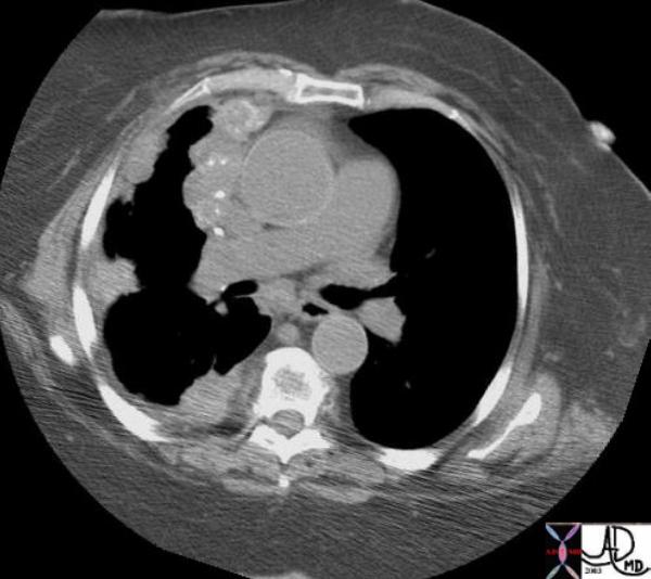
Pericardial Metastases from Primary Ovarian Carcinoma |
| A soft tissue density mass with punctate calcifications associated with pleural masses and thickening are noted on this chest CT. The patient has metastatic ovarian carcinoma to the pleura and pericardium with calcification. Courtesy Ashley Davidoff MD.
16864 Courtesy Ashley Davidoff Copyright 2009 |
|
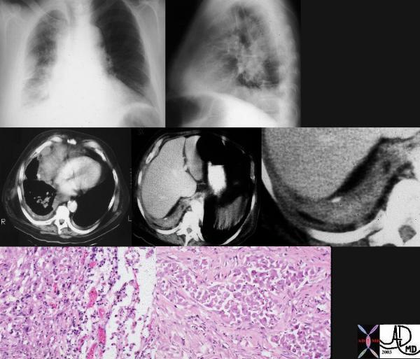
Pericardial Involvement with Malignant Mesothelioma
|
| This combination of images are from a patient with mesothelioma associated with asbestos related disease. Note the pleural plaques in images 3,4,5, the contracted hemithorax (1), the pleural origin of the density (2) characterised by blunted costophrenic angle, and the rind of soft tissue around the heart (3) and in the posterior costophrenic spavce. (3,4). The pathology shows a combination of malignant proliferation of stromal and glandular elements (7) abutting relatively normal lung (6)
Courtesy Ashley Davidoff MD. 32215c |
|
Lymphoma |
| 76349b03 heart pericardium fluiud hyperemic pericardial effusion lymphoma Courtesy Ashley Davidoff MD |
Links and References
Napolitano G et al Imaging Features of Constrictive Pericarditis: Beyond Pericardial Thickening Canadian Association of Radiologists Journal Volume 60, Issue 1, February 2009, (good review)
Senapati A et al, Disparity in spatial distribution of pericardial calcifications in constrictive pericarditis Openheart BMJmj. Volume 5, Issue 2
Khalid, N et al Pericardial Calcification StatPearls