
Hypertrophic Cardiomyopathy
Copyright 2020
Atlas of Cases
|
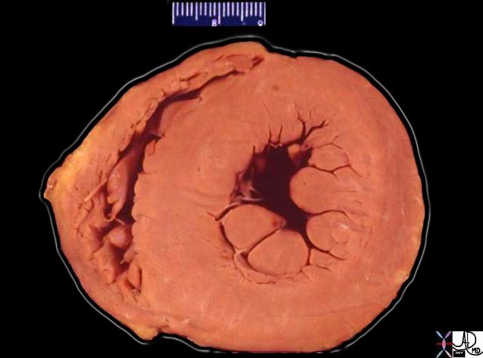
Concentric Hypertrophy |
| 08028b06 heart cardiac LV left ventricle papillary muscles RV right ventricle LVH left ventricular hypertrophy thickened LV concentric hypertrophy interventricular septum thickened enlarged grosspathology Courtesy Ashley Davidoff MD |
|
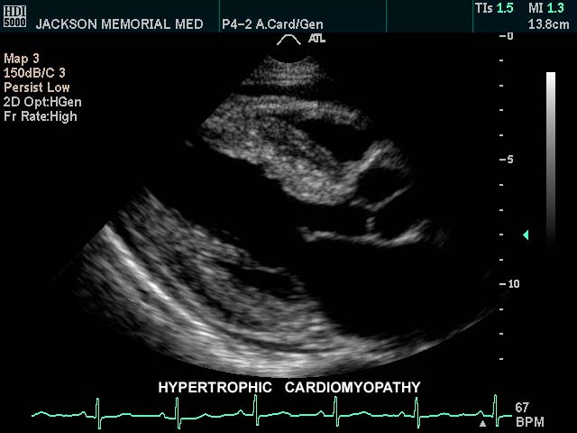
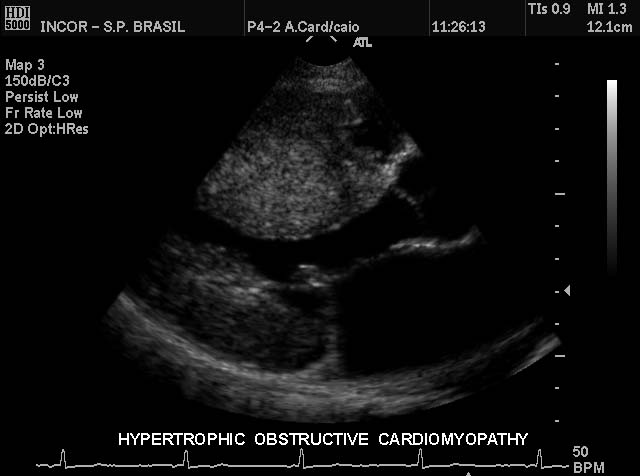
Hypertrophic Cardiomyopathy |
| This gray scale echo of the heart shows the left ventricle, anterior and posterior leaflets of the mitral valve, the aortic valve and the base of the aorta. There is a focal thickening of the ventricular septum in the left ventricular outflow tract just proximal to the aortic valve. The region is also slightly more echogenic than the remaining myocardium. This case demonstates a case of asymmetric septal hypertrophy or muscular subaortic stenosis. Courtesy Philips Medical Systems 33134 code cardiac heart echo LV MV ASH HOCM [dromano] |
|
IHSS |
| 33134c04.8s This gray scale echo of the heart shows the left ventricle, anterior and posterior leaflets of the mitral valve, the aortic valve and the base of the aorta. There is a focal thickening of the ventricular septum in the left ventricular outflow tract just proximal to the aortic valve. The region is also slightly more echogenic than the remaining myocardium. This case demonstates a case of asymmetric septal hypertrophy or muscular subaortic stenosis. Courtesy Philips Medical Systems 33134 code cardiac heart echo LV MV ASH HOCM |
|
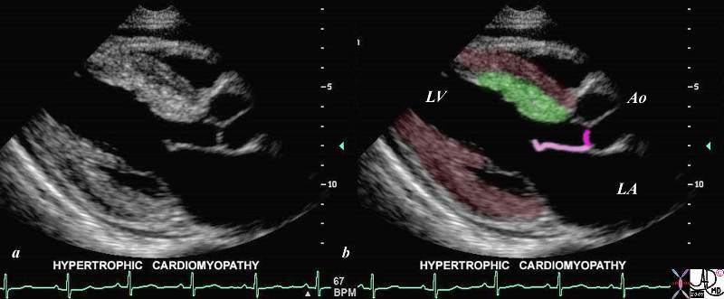
IHSS |
| This is a case of focal thickening (bright red overlay) in the region of the septum, which causes obstruction of the LV outflow tract during systole. This condition is known as asymmetric septal hypertrophy – also known as idiopathic hypertrophic sub-aortic stenosis -IHSS. Courtesy of Philips Medical Systems, Ultrasound, and modified by Ashley Davidoff M.D. 32133 |
|
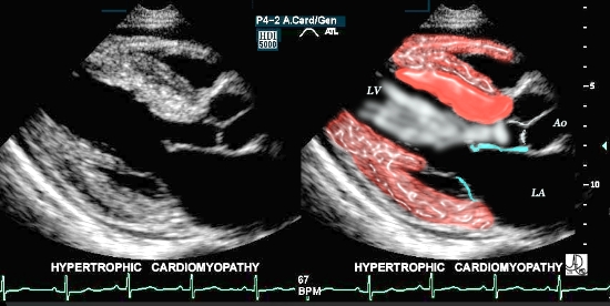
IHSS |
| This gray scale echo of the heart shows the left ventricle, anterior and posterior leaflets of the mitral valve, the aortic valve and the base of the aorta. There is a focal thickening of the ventricular septum in the left ventricular outflow tract just proximal to the aortic valve. The region is also slightly more echogenic than the remaining myocardium. This case demonstates a case of asymmetric septal hypertrophy or muscular subaortic stenosis. Courtesy Philips Medical Systems 33135 code cardiac heart echo LV septum thick LVOT narrow echogenic ASH IHSS imaging cardiac echo |
|
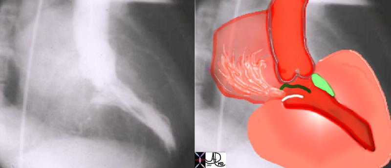
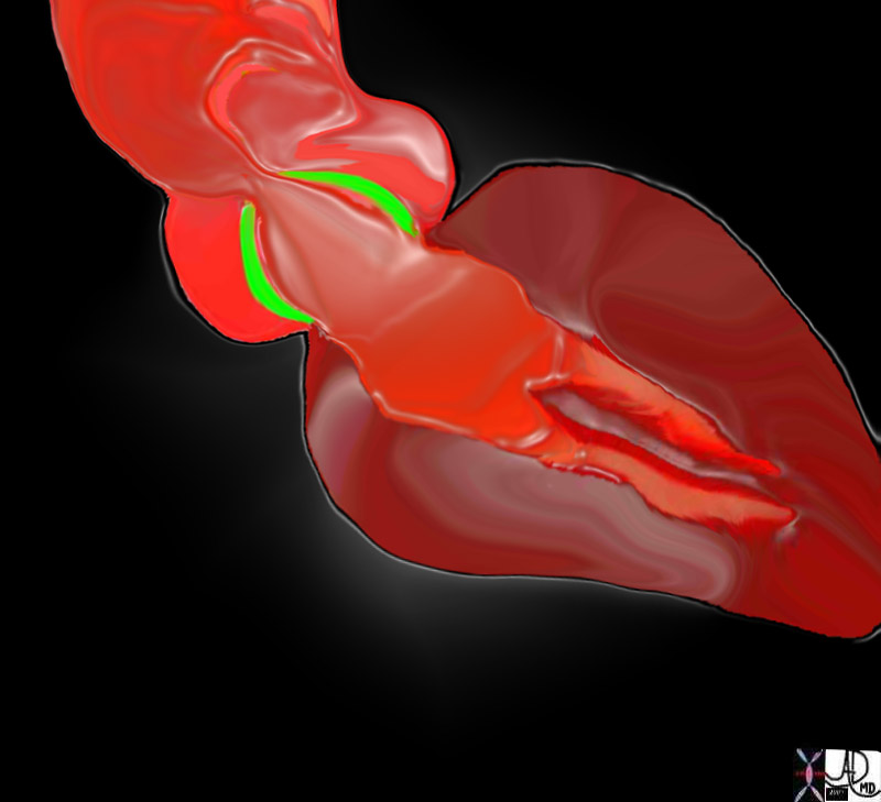
IHSS Angiography |
| This angiogram in RAO projection shows a hypercontractile left ventricle that has a ballet shoe appearance, with mitral regurgitation filling the left atrium. The drawing shows the significant LVH small cavity of the LV, the area of subaortic muscle bundle (green) and the mitral regurgitation caused by the systolic anterior motion of the mitral valve.Courtesy Ashley Davidoff 34805 cardiac heart MV interventriclar septum LVH IHSS SAM MR imaging radiology angiography disease overlay |
|
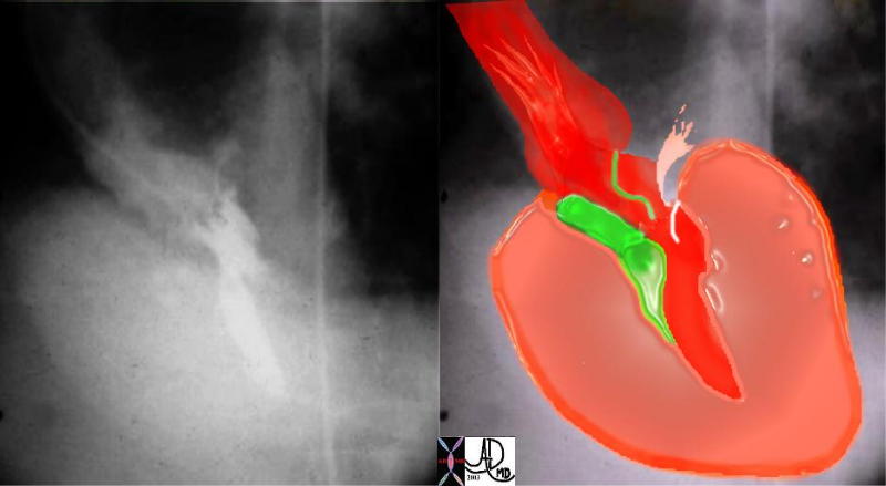
IHSS – RAO Projection |
| This angiogram in LAO projection shows a hypercontractile left ventricle that has a ballet shoe appearance, with mitral regurgitation filling the left atrium. The drawing shows the significant LVH, small cavity of the LV, the area of subaortic muscle bundle (green) and the mitral regurgitation caused by the systolic anterior motion of the mitral valve.Courtesy Ashley DAvidoff MD 34806 cardiac heart MV interventriclar septum LVH IHSS SAM MR imaging radiology angiography disease overlay |
|
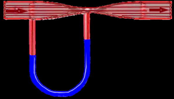
Venturi Effect |
| Flow in this tube is demonstrated by the arrows going from right to left through a narrowingin the tube (red) demonstrating a vacuum or suction effect caused by the sudden acceleration of the fluid as it goes through the narrowing or stenosis. This is seen in the U shaped monometer with the fluid level in blue being relatively higher at the site of the stenosis in relation to the pressure more upstream. The suction phenomenon is kown as the Venturi effect is seen in IHSS where the narrowing of the LVOT causes a vacuum effect on the anterior leaflet of the MV resulting in mitral regurgitation. The Venturi effect is also utilised in the functioning of carburettors in fuel driven engines.34807b05 code heart cardiac SAM IHSS pressure drop vacuum suction acceleration stnosis Venturi effect tube principles |
|
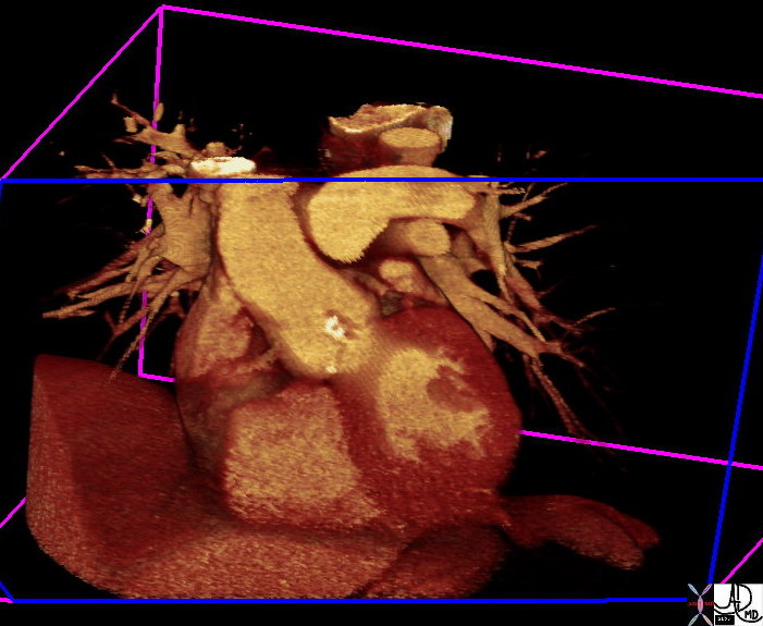
Aortic Stenosis |
| 39699.800 aorta aortic valvaortic annulus calcified calcification aortic sclerosis aortic stenosis LVH left ventricle hypertrophy CTscan volume rendering Courtsey Ashley Davidoff MD |
|
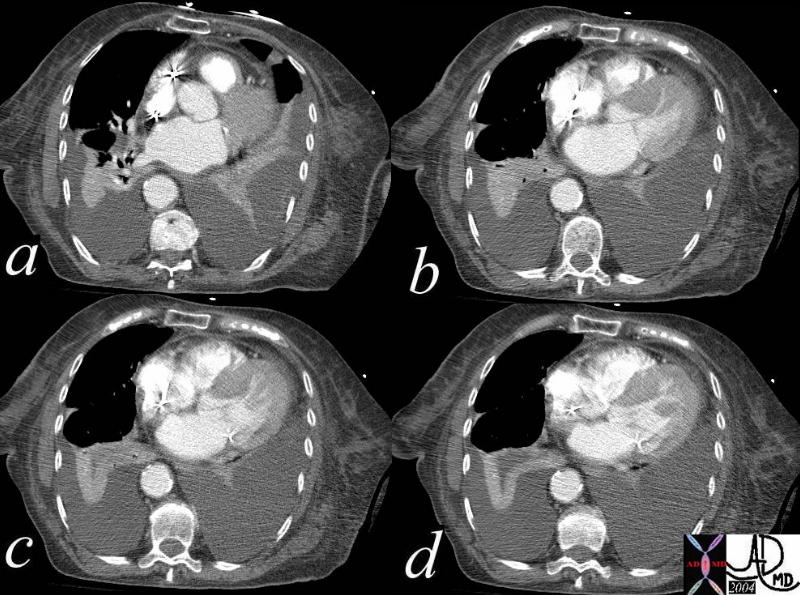
IHSS |
| This series of axial CTscans show asymmetric thickening of the ventricular septum in this patient with known diagnosis of IHSS (idiopathic hypertrophic subaortic steosis) and known systolic anterior motion of the mitral valve. The CT was performed one day after an RV perforation following placement of a pacemaker. Note also the left atrial enlargement, thickening of the RVOT, bilateral pleural effusions with passive compressive atelectasis. Courtesy Ashley Davidoff MD 39203c code CVS heart LV myocardiun thickness asymmetric hypertrophy RS lung pleura effusion atelectatic imaging radiology CTscan |
|
Aortic Stenosis |
| 07969bW.802 heart cardiac aorta aortic valve fx thickening of the aortic valves LVH left ventricular hypertrophy post stenotic dilatation of the ascending aorta turbulence eccentric jet doming of the aortic valve AV AS aortic stenosis Davidoff art |
|
16906.800 |
| 16906.800 heart LV left ventricle aorta coarctation left ventricular hyperrophy fx LVH concentric hypertrophy dx coarctation imaging radiology T1 weighted MRI Courtesy Ashley Davidoff MD |
|
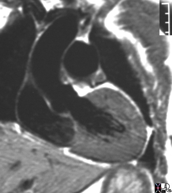
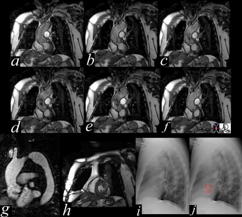
38871c01 |
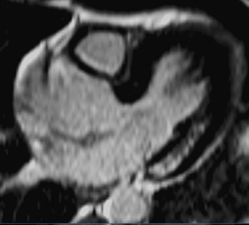
| This series of coronal MRI images of the aortic valve (a-f) show phases from diastole (a) through systole (b,c,d,e) with a narrow (b,c) and then turbulent jet, (d,e) back to diastole (f) Image g shows a thickened valve, while the short axis of the LV (h) shows LV hypertrophy. The plain film of h and i highlight the calcific nature of the valve. The diagnosis is aortic valve stenosis. Courtesy Scott TSai MD 38871c01 code cardiac heart aortic valve AS LVH calcification calcified imaging radiology MRI CXR plain film |
|
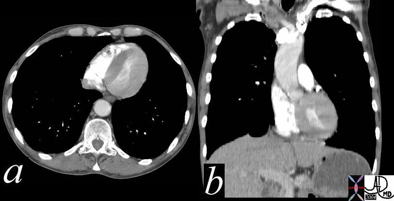
39088c01 |
| Coronal reformats through the chests of two different patients. They are approximately at the same level of the RVOT. The left image (a) is normal, while image b shows concentric thickening of the LV. Courtesy Ashley Davidoff MD 39088c01 code cardiac heart normal LVH LV hypertrophy |
|
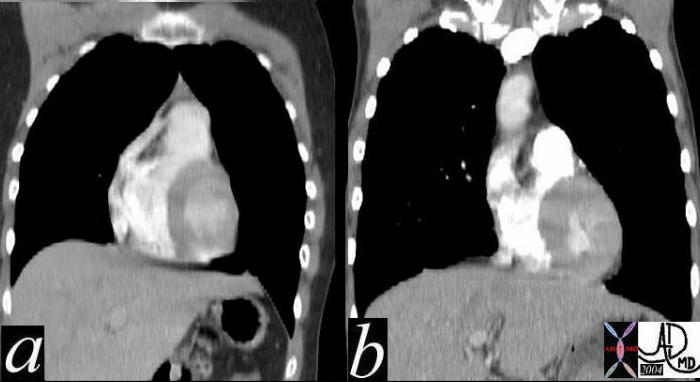
39088c |
| This axial and coronaaly reformatted CTscan of the left ventricle and the aorta show concentric hypertrophy of the LV and ectasia of the ascending aorta in this patient with severe systemic hypertension. Courtesy Ashley Davidoff MD. 39088c code CVS LV LVH AO ectasia hypertension |
|
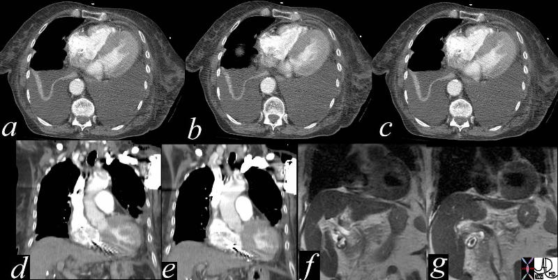
39221c |
| This series of axial and coronal CT scans (a,b,c,d,e) and coronal T1 weighted MRI scans show symmetric thickening of the left ventricle in this patient with LV hypertrophy. The MRI preceded the placement of the pacemaker noted in d and e. Courtesy Ashley Davidoff MD 39221c code cardiac heart LV myocardium thick concentric hypertrophy imaging radiology CTscan |
Fabry Disease
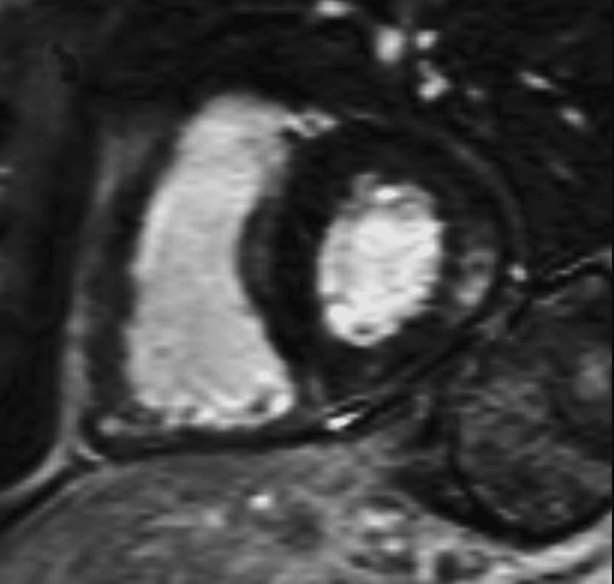
“Delayed enhanced images of 64-year-old heterozygotic woman with Fabry’s disease–related cardiac hypertrophy. Minimal thinning of basal segment of left ventricular inferolateral wall is associated with thick mesocardial striae of delayed enhancement shown on short-axis delayed enhanced images.”
De Cobelli et al Delayed-Enhanced Cardiac MRI for Differentiation of Fabry’s Disease from Symmetric Hypertrophic Cardiomyopathy
AJR Volume 192, Issue 3 2009
“40-year-old man with Fabry’s disease–related hypertrophy. Long-axis delayed enhanced images show typical pattern of delayed enhancement: thick striae involving inferolateral wall of basal segment of left ventricle in mesocardial distribution. Note sparing of subendocardial layer.
De Cobelli et al Delayed-Enhanced Cardiac MRI for Differentiation of Fabry’s Disease from Symmetric Hypertrophic Cardiomyopathy”
AJR Volume 192, Issue 3 2009
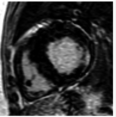
 LGE in FABRY DISEASE
“40-year-old man with Fabry’s disease–related hypertrophy. Short -axis delayed enhanced images show typical pattern of delayed enhancement: thick striae involving inferolateral wall of basal segment of left ventricle in mesocardial distribution. Note sparing of subendocardial layer.”
De Cobelli et al Delayed-Enhanced Cardiac MRI for Differentiation of Fabry’s Disease from Symmetric Hypertrophic Cardiomyopathy”
AJR Volume 192, Issue 3 2009
TCV Cases
References and Links
- TCV