
Copyright 2008
Dima Quraini MD Ashley Davidoff MD
Definition
Pericarditis is an inflammatory or infectious disorder of the pericardium and has a multitude of causes. It is commonly encountered in patients inflicted with autoimmune diseases such as rheumatoid arthritis or lupus, patients with uremia and patients who have undergone cardiac surgery. Pericarditis may also result from a viral, bacterial or fungal infection.
Patients presenting with bacterial pericarditis tend to be very sick and usually present with fever and sepsis rather than isolated chest pain. Myocardial ischemia or infarction can result in inflammation of the pericardium either acutely or a few weeks after the acute event. Dressler’s syndrome is a specific immune syndrome consisting of fever and pericarditis occurring weeks after a myocardial infarction. The incidence of ischemia or infarct related pericarditis has declined since the advent of early coronary revascularization and the use of thrombolytics.
Acute pericarditis results in a serositis so that the inflammed pericardium presents with a somatic pain, and also exudes fluid into the pericardial space.
Acute pericarditis can be complicated by pericardial tamponade if there is a rapid accumulation of pericardial fluid.
Clinical presentation includes central, pleuritic chest pain that, as a result of the pathway of the phrenic nerves, can radiate to one or both trapezius ridges. The pain may be aggravated by breathing simulating pleuritic pain or when the patient lies down. It is often relieved when sitting up and leaning forward. Pain may worsen when patients lie down and may improve when they sit up and lean forward. On auscultation of the heart, a monophasic, diphasic or triphasic pericardial rub can be appreciated. Pericardial rubs are highly specific for pericarditis however require careful or skillful auscultation to be picked up. Their low sensitivity is also due to their fleeting or intermittent nature.
The diagnosis is suspected by the classical clinical presentation, and larger pericardial effusions may cause decrease EKG voltages. However the diagnosis is best confirmed with echocardiography
Treatment includes anti-inflammatory drugs such as aspirin, NSAID or prednisone. Aspirin is effective; however the mostly utilized treatment for pericarditis is ibuprofen. It can be combined with colchicine that can help prevent recurrent episodes of pericarditis. The combination therapy should last 7 to 14 days then tapered over a week or two to prevent recurrence. Oral prednisone is used in patients with severe pericarditis and who do not respond to NSAIDs.
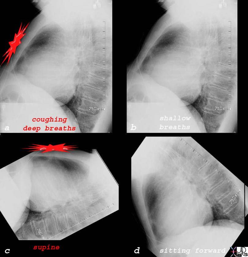
Aggravating and Relieving Characteristics of Pain from Pericarditis |
| 76031c01d02 chest heart pericardium pain aggravated by coughing and deep breathing relieved by shallow breathing and sitting forward acute pericarditis pericardial effusion lateral chest X-ray plain X-ray Courtesy Ashley Davidoff MD |
On auscultation of the heart, a monophasic, diphasic or triphasic pericardial rub can be appreciated. Pericardial rubs are highly specific for pericarditis however require careful or skillful auscultation to be picked up. Their low sensitivity is also due to their fleeting or intermittent nature.
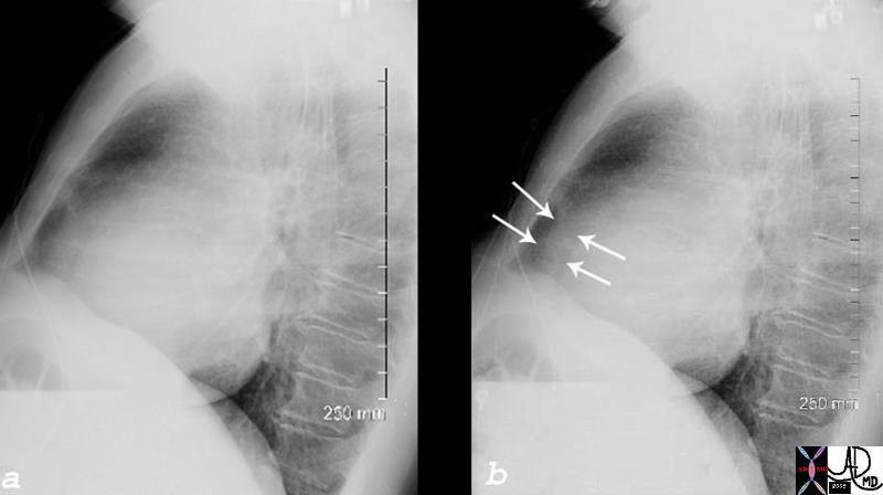
Lateral Chest Examination Showing Pericardial Effusion |
| 76031c01d04 chest heart pericardium aggravated by coughing and deep acute pericarditis pericardial effusion lateral chest X-ray plain X-ray Courtesy Ashley Davidoff MD |
Acute Pericarditis |
| 76031c01d heart cardiac pericardium pericardial effusion enlarged fluid between two fat layers sandwich sign visceral pericardium parietal pericardium epicardial fat pericardial fat plain film CXR plain X-ray Courtesy Rebecca Schwartz |
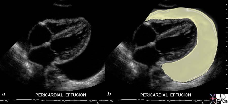
Large Pericardial Effusion – Echocardiogram |
| 33140c01 This gray scale echo of the heart showing a 4 chamber view, and demonstrating a large collection of fluid around the heart, characteristic of a pericardial effusion. Courtesy Philips Medical Systems 33140 code cardiac heart echo pericardium effusion imaging cardiac echo overlay Davidoff MD |
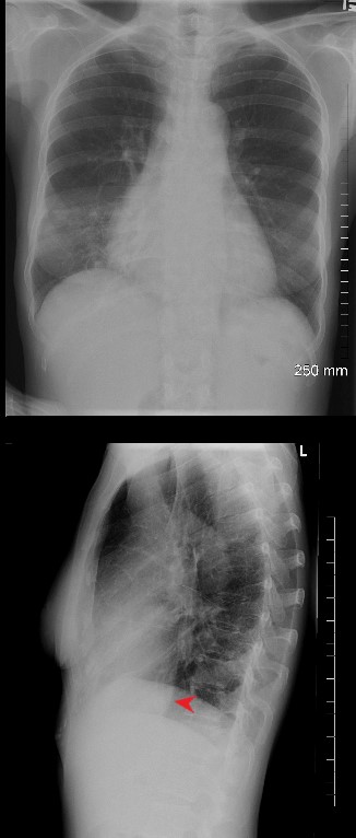
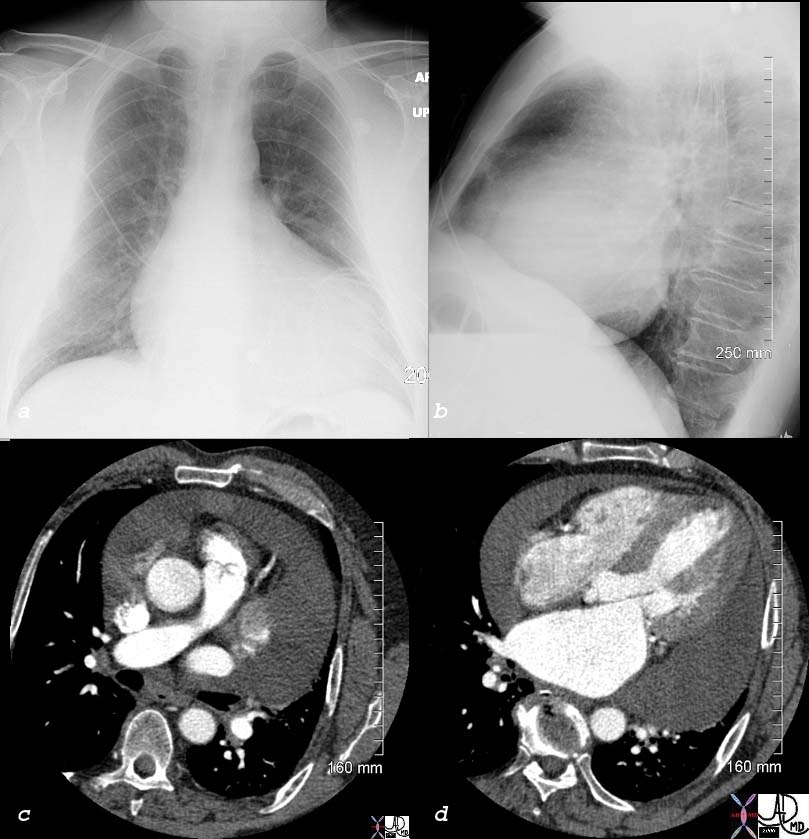
2 views of the chest of a 54 year old female with SLE and Sjogren’s syndrome show a slightly enlarged heart in the PA projection and an unusually prominent and rotund IVC (red arrowhead)
Ashley Davidoff MD
key words
pericarditis, pericardial effusion, SLE
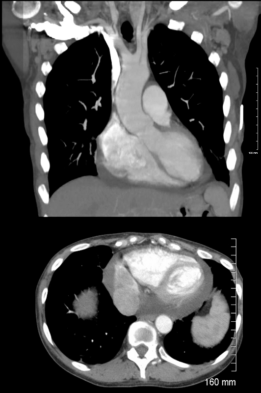
Coronal (above) and axial images CT showing the Rv and LV of a 54 year old female with SLE and Sjogren’s syndrome. A small pericardial effusion is noted accounting for the cardiomegaly on CXR.
Ashley Davidoff MD
key words SLE, pericarditis, pericardial effusion