
Ashley Davidoff MD
Definition
The left ventricular outflow tract (LVOT)is the part of the left ventricle adjacent to the aortic opening. It is also called the aortic vestibule; the aortic vestibule differs from the rest of the ventricle in that it has fibrous and not muscular walls.
Structurally, the LVOT is made up of the anterior leaflet of the mitral valve positioned on the left wall and boundary, and medially the ventricular septum, and membranous septum act as the other boundary. The bundle of His divides in the mebranous septum and travels through the septum and down the muscular septum so that it travels through the medial wall of the LVOT.
Functionally the LVOT receives blood during systole when the anterior leaflet of the mitral valve is pushed leftward by the contractile forces exerted on it and acts as a conduit for the blood to be transported to the aorta via the aortic valve. During diastoles it narrows because the anterior leaflet is opened by diatolic flow from the left atrium to the left ventricle.
Diseases of the LVOT include diseases of the anterior leaflet of the mitral valve, ventricular septum, and membranous septum. A cleft mitral valve for example is a maloriented split valve and it causes narrowing and elongation of the LVOT during diatole manifesting as a gooseneck deformity on angiography. SAM is a classical deformity of the LVOT during systole in IHSS that results in mitral reurgitation.
Diagnosis of diseases in the LVOT are best performed by echocardiography but MROI and CT can augment echo.
Treatment is based on the disease and may be surgical or medical.
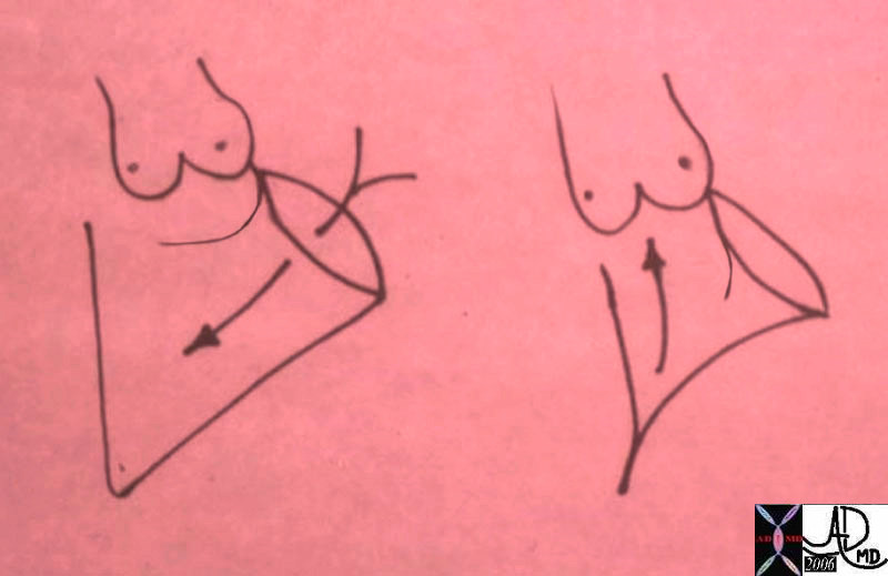
Inflow and Outflow of the Left Ventricle |
| The mitral valve participates in the inflow as well as the outflow of the LV. The LV does not have an infundibular chamber like the RV. It is the anterior leaflet of the MV that has this dual function. The first drawing represents LV diastole, showing the open anterior leaflet acting as the anterior, medial and rightward border of the inflow to the LV. The second drawing is the systolic phase where this same anterior leaflet acts as the leftward and lateral border of the outflow tract.
Courtesy of Ashley Davidoff M.D. 32119b01 |
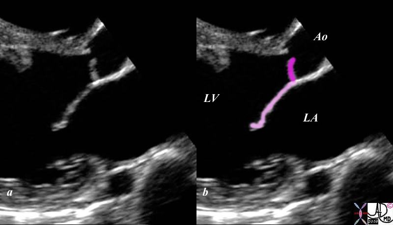
The Normal Mitral Aortic Complex |
| This gray scale echo of the heart shows the left ventricle, anterior and posterior leaflets of the mitral valve, the aortic valve and the base of the aorta. The echo demonstrates the intimate anatomic and physiological relationship of the anterior leaflet of the mitral valve (light pink) and the left aortic cusp to best effect.
( Courtesy Philips Medical Systems 33132 code cardiac heart echo LV MV aorta mitralaortic complex normal anatomy 33132c02.8s |
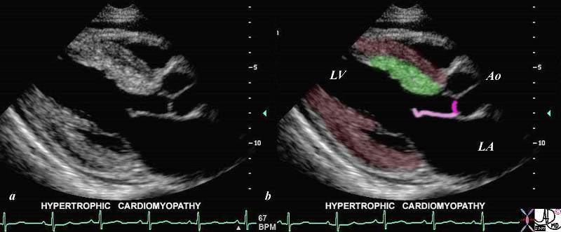
SAM and IHSS |
| This gray scale echo of the heart shows the left ventricle, anterior and posterior leaflets of the mitral valve, the aortic valve and the base of the aorta. There is a focal thickening of the ventricular septum in the left ventricular outflow tract just proximal to the aortic valve. The region is also slightly more echogenic than the remaining myocardium. This case demonstates a case of asymmetric septal hypertrophy or muscular subaortic stenosis.
Courtesy Philips Medical Systems 33134 code cardiac heart echo LV MV ASH HOCM 33134c04.8s |
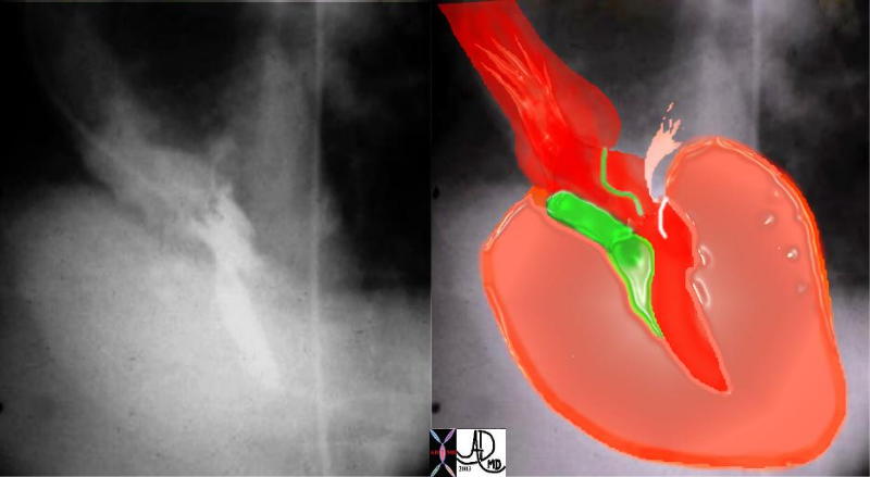
Narrowing of LVOT – Subaortic Stenosis – IHSS |
| This angiogram in LAO projection shows a hypercontractile left ventricle that has a ballet shoe appearance, with mitral regurgitation filling the left atrium. The drawing shows the significant LVH, small cavity of the LV, the area of subaortic muscle bundle (green) and the mitral regurgitation caused by the systolic anterior motion of the mitral valve. Courtesy Ashley DAvidoff MD 34806 cardiac heart MV interventriclar septum LVH IHSS SAM MR imaging radiology angiography disease overlay |

Aortic Regurgitation and the Austin Flint Murmur |
| This gray scale echo of the heart showing a right parasternal long-axis left ventricular outflow view, and demonstrating prolapse of the aortic valve. The patient has a diagnosis of aortic regurgitation. The regurgitant jet of the aortic incompetence created by the prolapsing valve causes the mitral leaflet to flutter..
Courtesy Philips Medical Systems 33124 cardiac heart echo AOV prolapse MV imaging echo 33124c02.8s |
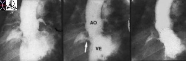
LVOT VSD of the Membranous Septum |
| This is a series of angiograms of the left ventricle in LAO projection showing a puff of contrast into the right ventricle through the interventricular septum. A similar collection of contrast is noted in the the main pulmonary artery just to the left of the aorta. This high VSD is in the position of the membranous septum and thus represents a mebranous ventricular septal defect.
Courtesy Ashley Davidoff MD. 00250 code cardiac heart LV RV VSD membranous MPA aorta AO congenital imaging radiology angiography |