
69 year old male presented with history of cardiomyopathy and atrial fibrillation
The LGE on the short axis shows biventricular thickening of the RV and LV
There are other findings on the MRI that are highly suggestive of sarcoidosis. There are multicentric foci of LGE in linear and nodular form in the mid myocardial and subepicardial layers and likely in the pericardium and myocardium of the right ventricle.
There was associated global hypokinesis of the LV with an EF of 40%, and increase in the LV mass of 120gms/ sq m
Ashley Davidoff MD
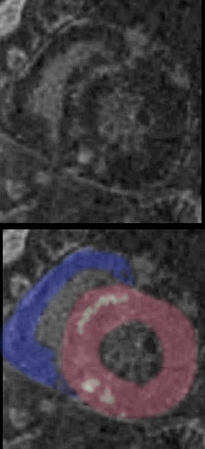
48-year-old man with a previous history of sudden loss of vision which subsequently resolved and presents now with near syncope, atrial fibrillation. MRI showed multifocal acute embolic infarcts involving the cerebellum. Echocardiogram showed an enlarged left atrium, significant left ventricular hypertrophy, and moderate mitral regurgitation.
CXR shows LAE and LVE with cephalisation
The MRI shows multifocal linear and nodular LGE at the hinge points (green) in the subepicardial regions (blue) at the base of the heart anteriorly and inferiorly, as well as mid myocardial linear LGE in the septum (pink). The bright LGE is dominant in the nodular form (red)
Diffuse hypokinesis, LVH (95gms/sq m) EF of 39%, moderate mitral regurgitation, and significant left atrial enlargement
Sarcoidosis is though to be most likely together with nonspecific hypertrophic cardiomyopathy . Distribution of LGE is not characteristic of Fabry disease. Amyloidosis is also considered as a less likely possibility
Ashley Davidoff MD

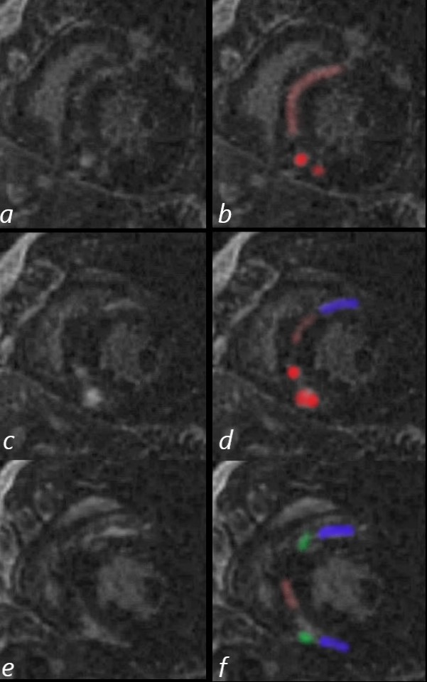
69 year old male presented with history of cardiomyopathy and atrial fibrillation
The findings on MRI are highly suggestive of sarcoidosis. There are multicentric foci of LGE in linear and nodular form in the mid myocardial and subepicardial layers and likely in the pericardium and myocardium of the right ventricle.
In this series images on the right (b,,d,,f) are magnified views of images on the left (a,c,e)
There are multicentric foci of LGE nodular form in the mid myocardial region (white arrowheads) linear LGE in the subepicardial layers (red arrowheads, b and f) in the pericardium (teal blue arrowhead) and myocardium of the right ventricle (yellow arrowhead e).
There was associated global hypokinesis of the LV with an EF of 40%, and increase in the LV mass of 120gms/ sq m
Ashley Davidoff MD
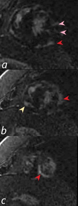
69-year-old male presented with history of cardiomyopathy and atrial fibrillation
There are multicentric foci of LGE in this short axis view taken closer to the apex with linear form in the mid myocardial region (pink arrowheads) and linear and nodular changes of LGE in the subepicardial layers (red arrowheads, a,b,c) Subepicardial changes are also notes in the RV in a (not marked)
The findings on MRI are highly suggestive of sarcoidosis.
There was associated global hypokinesis of the LV with an EF of 40%, and increase in the LV mass of 120gms/ sq m
Ashley Davidoff MD

69 year old male presented with history of cardiomyopathy and atrial fibrillation
In this series the long axis 4 chambered view reveals intense continuous mid myocardial LGE
Ashley Davidoff MD
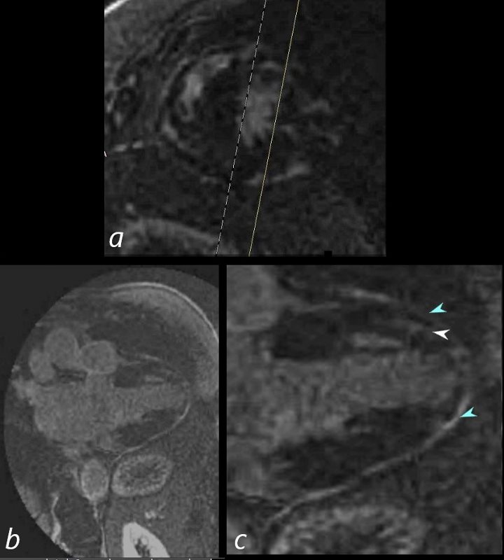
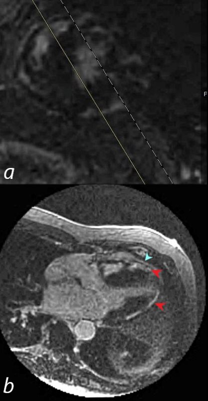
69-year-old male presented with history of cardiomyopathy and atrial fibrillation
In this series, the long axis 2 chambered view (c is magnified view of b) reveals intense continuous pericardial disease (teal blue arrowheads) and mid myocardial linear-nodular LGE (white arrowhead)
Ashley Davidoff MD
69-year-old male presented with history of cardiomyopathy and atrial fibrillation
In this series the long axis 3 chambered view reveals intense continuous sub epicardial disease (red arrowheads) and pericardial LE (teal blue arrowhead)
Ashley Davidoff MD