
Copyright 2009
Definition
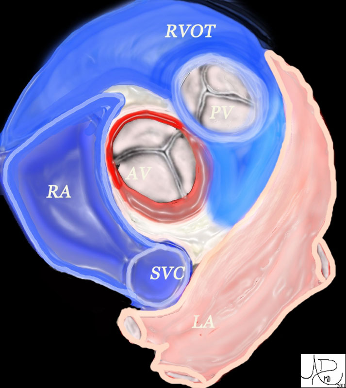
The pulmonary valve is the semilunar valve that guards the opening of the right ventricular outflow tract into the pulmonary artery. Structurally the pulmonary root, which forms the basic functional unit of the valve includes the pulmonary annulus, sinuses of Valsalva, the valvular leaflets, muscular infundibulum and the sinotubular ridge. Functionally, by passive opening and closing secondary to pressure changes on either side of the valve , it serves to transmit blood in one direction from the RV to the pulmonary artery and not vice versa .
Primary Diseases of the pulmonary valve are not very frequent but, are often a accompaniment of most congenital heart diseases.
Diagnosis of pulmonary valve stenosis or regurgitation is suspected in patients presenting with dyspnea, chest pain, syncope, dizziness and a murmur in the pulmonary area with a loud and variable or fixed splitting of the second heart sound S2. Chest radiography may show pulmonary artery dilation or increased vascularity of the peripheral lung fields or oligemia depending on the valve disease. EKG shows RV hypertrophy with right axis deviation. Echocardiography as in all valve disease allows for assessment of valve anatomy and function.
Medical therapy for congestive heart failure or infective endocarditis, minimally invasive techniques and surgical options are available.
Structural Considerations:
The pulmonary valve complex includes the pulmonary annulus, sinuses of Valsalva, the valvular leaflets, muscular infundibulum and the sinotubular ridge. The pulmonary valve is the most superiorly placed valve.
The sinotubular junction separates the pulmonary valvular sinuses from the tubular component of the pulmonary trunk. Muscular infundibulum or conus refers to the smooth muscular out flow portion of the RV.The sinuses of Valsalva are the three parts of the pulmonary root confined proximally by the semilunar attachments of the valvular leaflets and distally by the sinotubular junction. The valve leaflets are three in number and are named as right, left and non septal cusp (anterior). The pulmonary valve has no true annulus but there are fibrous semilunar attachments which support the cusp.
The pulmonary valve area of a healthy adult is 2.0 cm2/m2 of body surface area.
|
47681 |
| 47681 heart cardiac aortic valve aorta right atrium right atrial appendage left atrial appendage LAA RAA pulmonary valve right ventricular outflow tract left atrium LA RA RVOT anatomy normal position relations Davidoff art Davidoff MD |
|
00272c |
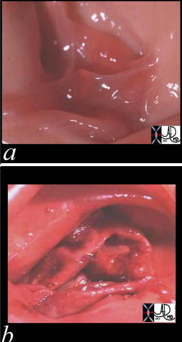
| These two pathological specimens of the pulmonary valve show the normal delicate leaflets of the normal valve (a) in comparison to the thickened leaflets of the bicuspid pulmonary valve Courtesy Ashley Davidoff MD 00272c code CVS PA MPA PV normal bicuspid pulmonary valve stenosis |
|
01473c02 |
| This pathological specimen shows an anteriorly positioned larger aorta (bright red in b)in front and to the right of the the string like atretic , posteriorly placed main pulmonary artery (royal blue in b). The ductus arteriosus (green overlay in b) is one of the mechanism via which the pulmonary circulation is able to maintain flow. Courtesy Ashley Davidoff MD 01473c02 code heart pulmonary atresia D transposition LTGV LTGA congenital grosspathology |
01483c |
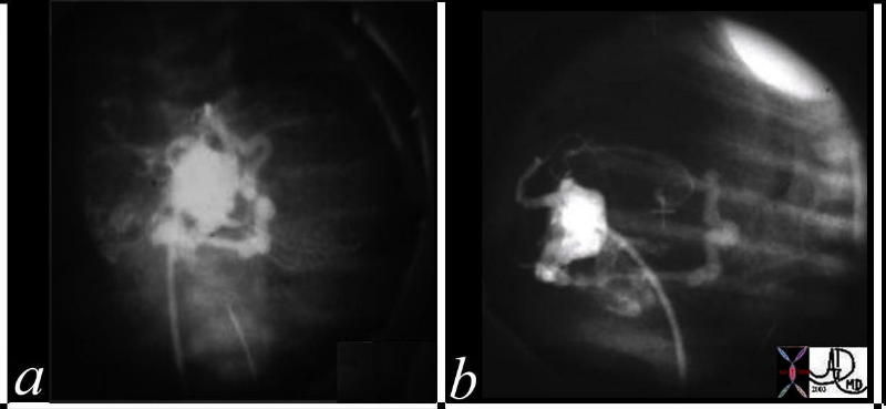
| This is an RV angiogram of a neonate with hypoplastic right heart syndrome. The catheter course from the IVC to the RA and then the RV. The tricuspid valve was barely open and there was no egress for blood via the RVOT which was atretic. The tubular structures seen in a and b are the coronary veins and arteries that are filling in retrograde fashion, from the isolated RV through the venous channels that drain directly from the RV into the cardiac veins. From the veins they pass into the arteries which are faintly seen in the upper portions of b. (lateral projection) Courtesy Ashley
Davidoff MD 01483c code CVS heart cardiac coronary artery pulmonary atresia hypoplastic right heart syndrome retrograde filling of coronary arteries veins imaging radiology angiography |
|
00274c02 |
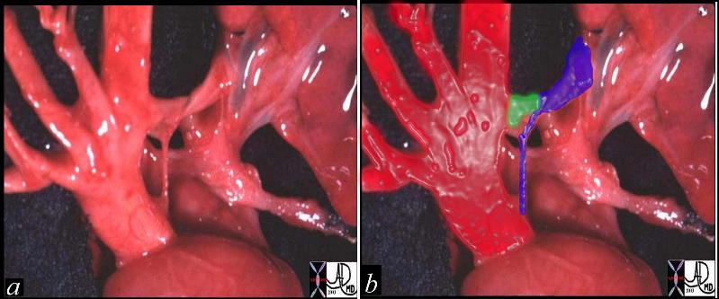
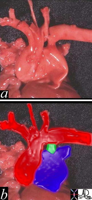
| This post mortem specimen reveals the great vessels with the aorta overlaid in red and the pulmonary artery overlaid in blue (b). The MPA is dilated due to an underlying congenital pulmonary valve stenosis (PS) The green overlay is a patent ductus arteriosus (PDA) Courtesy Ashley Davidoff MD 00274c02 see also 00274c04 |
|
00274c04 |
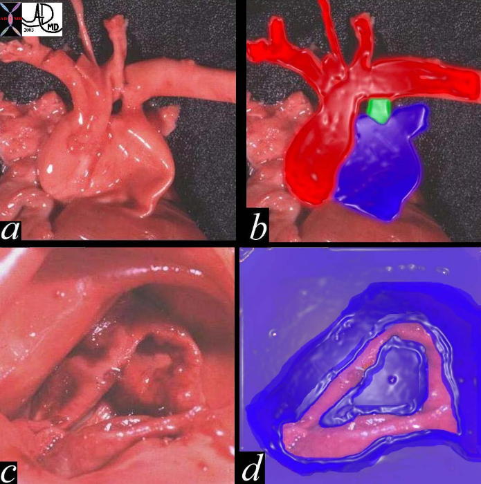
| This post mortem specimen reveals the great vessels with the aorta overlaid in red and the pulmonary artery overlaid in blue (a, b). The MPA is dilated due to an underlying congenital bicuspid pulmonary valve resulting in stenosis (PS) The green overlay (b) represents a patent ductus arteriosus (PDA) In image c and d the thickened pulmonary valve is identified with fusion of the commissures to form a bicuspid stenotic valve. In image d the valve is outlined in pink fleshy color. Courtesy Ashley Davidoff MD 0400274c04 see also 00274c02 |
|
15036c01 |
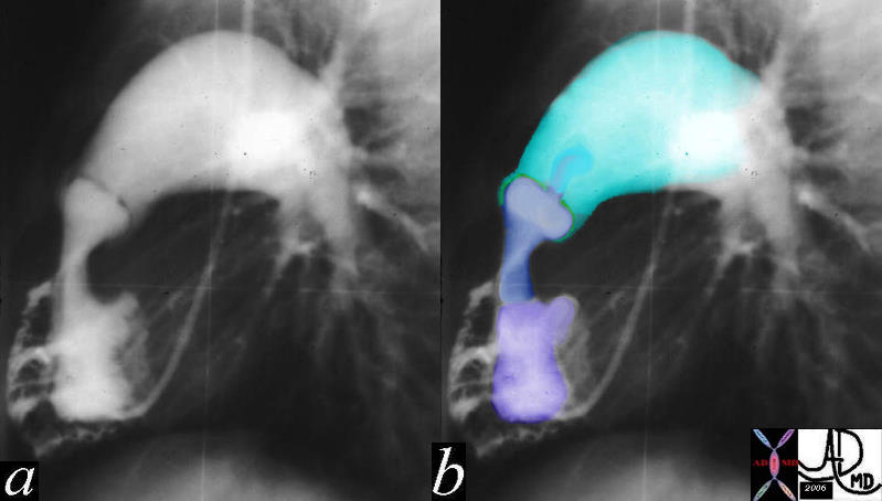
| 15036c01 lung pulmonary artery heart cardiac hypoplastic right ventricular syndrome hypoplastic right ventricle small right ventricle small RV PS pulmonary valve pulmonary stenosis post stenotic dilatation doming pulmonary valve intravasation into coronary arteries suprasystemic RV pressure congenital heart disease size shape RV angiogram angiography Davidoff MD |
Pulmonary Stenosis |
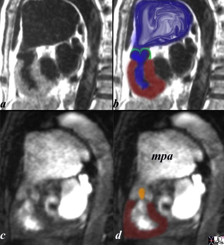
| The sagittal view using MRI with black blood (a,b) and white blood(c,d) imaging is from a patient with pulmonary stenosis. The RV (maroon overlay) is hypertrophied, the domed stenotic pulmonary valve (green overlay)is best seen in a and b, and the turbulence through the valve is seen in c, overlaid in orange in d. there is significant post stenotic dilatation of the main pulmonary artery (mpa) inferred by the turbulence in b (blue overlay).
Courtesy Ashley Davidoff copyright 2009 all rights reserved pulmonary valve 15437c03.8s |
Rheumatic Disease
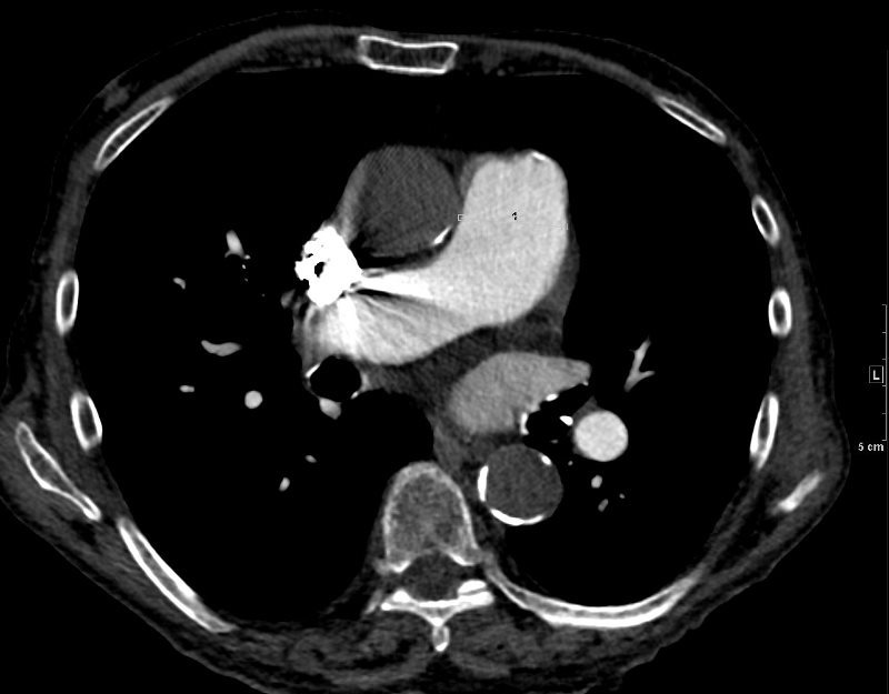
ENLARGED PA AND PV CALCIFICATION
87 year old male with findings consistent with mitral stenosis , characterised by calcified mitral valve, enlarged left atrium, normal sized left ventricle,with enlarged right atrium, right ventricle and main pulmonary artery.
Pure mitral stenosis is most commonly caused by rheumatic heart disease
Ashley Davidoff MD