
Foramen Ovale
pyright 2008
Definition
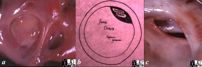
The foramen ovale is an crescentic shaped foramen that lies on the superior edge of the septum primum and acts as a flap valve in the fetus to allow blood to shunt from the right atrium to the left atrium.
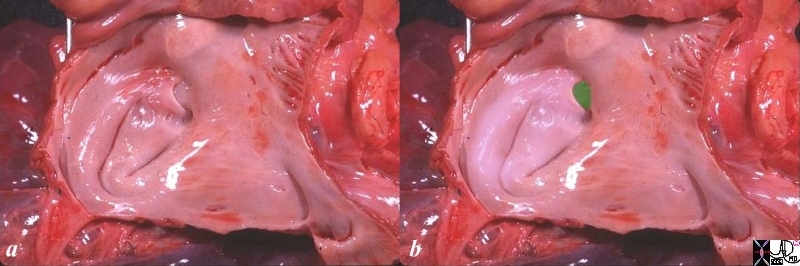
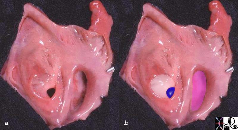
The left atrium does have curves to its inside surface but not to the extent of the right atrium, and it is mostly smooth. The septum primum (white) and foramen ovale (blue) are in the center. The mitral valve is overlaid in pink. Fine pectinate muscles are seen at the mouth of the finger like left atrial appendage.
Ashley Davidoff
TheCommonVein.net
Structurally its shape and position characterize it. More specifically it lies on the left atrial side and overlaps the superior limbic band of the septum secundum. When one looks at the atrial septum from the right atrium it is not visible in the normal situation. It is only seen from the left atrial side.
Embryologically it is felt to arise from the left venous valve of the sinus venosus, which is the precursor for the septum primum, upper portion of the atrial septum, svc, ivc, and the thebesian valves . (van Praagh)
Functionally in the fetus it plays a major role in acting as a conduit for the blood that is shunted from the right atrium to the left atrium so that nutrient filled blood from the mother can be directed to the fetal cerebral circulation and developing brain as well as the systemic circulation. In the fetus, the pulmonary resistance is high and therefore right atrial pressure is relatively high which causes the flap valve to shift toward rtthe LA and hence allows flow in that direction. At birth the first breat results in a drop of the pulmonary resistance and hence increases flow to the lungs, and thus LA pressure rises as the increasing volume of blood returns to the LA. The foramen ovale and surrounding tissue gets pushed against the superior limbic band and closes it functionally. Over time the fusion is consolidated and in 70% of people the closue is permanent. In 30% the closure is incomplete and a potential defect called a patent foramen ovale exists.
There are only a few diseases that affect the foramen ovale and include a patent formen oval and paradoxical embolus that may occur as a complication.
The diagnosis is usually clinically suspected whrn a patient shows manifestations of a paradoxical embolus, and is documented by echocardiography and particulalrly a “bubble” study.
Treatment if necessary is by minimal invasion or surgery.