Copyright 2009
Definition
Dextrocardia is defined as a malposition of the heart that can be congenital or acquired resulting in the apex of the heart being in the right chest. There are two congenital forms; One is associated with situs solitus, and the second associated with situs inversus. The form that is associated with situs solitus is thought to be due to an embryonic arrest resulting in the the right ventricle (rather than the left) forming the apex. In this instance the right ventricle is still on the right side and the left ventricle is on the left side. This form of dextrocardia is associated with a higher incidence of congenital abnormalities. The second form of the congenital version is associated with situs inversus. In this situation the incidence of associated congenital abnormalities is lower.
Acquired dextrocardia may be due to either loss of volume in the right chest or a push due to mass effect from the left side.

Dextrocardia |
|
The PA chest X-ray shows the apex pointing rightward and most of the heart is in the right chest. Note also that the stomach bubble is in normal position, and so situs solitus is present. This is a case of isolated dextrocardia, caused by an arrest of development, not associated with situs inversus, but subject to a higher incidence of other congenital anomalies. (Images courtesy of Ashley Davidoff M.D.) |
|
Dextrocardia with Situs Solitus |
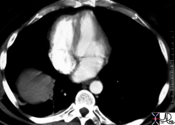
|
The CT is from an elderly patient with dextrocardia and situs solitus. The scout film before the CT shows dextrocardia and Harrington rods are noted in the thoracic spine. Image b, shows the apex of the heart pointing to the right, but less impressive than the plain film. Image c through the great vessels show normal relationship, and through the abdomen, situs solitus is confirmed. Courtesy Ashley Davidoff MD copyright 2009 45806c01.8s |
|
Dextrocardia and Situs Inversus |
|
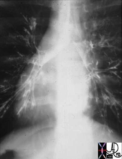
The CXR and bronchogram is from a 30year old male who presents with a long standing history of a productive cough, sinus headaches, and infertility. The bronchogram shows inversion of the bronchi with the long thin bronchus characteristic of the left bronchus being right sided and short “fat ” bronchus characteristic of the right being left sided. Situs inversus and dextrocardia exist. However the history of sinusitis and infertility make the diagnosis of Kartagener’s syndrome likely. 06248 Courtesy Ashley Davidoff Copyright 2009 all rights reserved |
|
Acquired Dextrocardia |
|
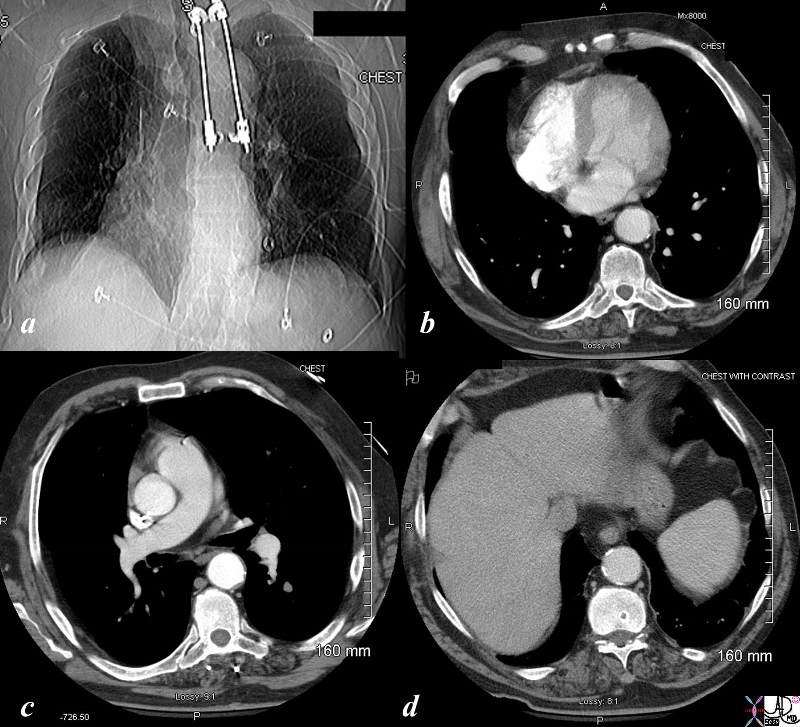
The series of CT scan show an acquired dextrocardia. In image a, the heart is not seen at all – but since it is not in the left chest, it is presumed to be in the right. The mediastinum as seen by the trachea is shifted to the right and the right lung is shifted across the midlineinto the left chest. the patient is status post pneumonectomy. Image b shows the heart completely in the right chest, and image c, shows the shift of the left lung across the midline and complete absence of the right lung. Image d confirms situs solitus Courtesy Ashley Davidoff copyright 2009 all rights reserved 86476c01.8s |
|
Spurious Dextrocardia |
|
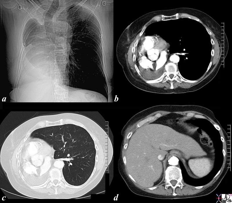
In this case the scout film manifests with what appears to be a case of dextrocardia. Image b shows a slight of the heart to the right but the appearance is dominated by by a large right atrium and left atrium. Image c confirms the leftward position of the apex and normal relationship of the left apex and right ventricle. Image d shows situs solitus with reflux of contrast into the dilated hepatic veins indicating tricuspid regurgitation. Courtesy Ashley Davidoff MD copyright 2009 48098c02 |

|
Mesocardia |
|
This is a CT scan through the chest in which the apex of the heart points forward. The left ventricle (LV) is left sided and the right ventricle is right sided. There is situs solitus of the atria. This is a case of mesocardia ie neither right nor leftwad pointing apex. Courtesy Ashley Davidoff MD. 19689 code heart mesocardia cardiac imaging radiology CTscan |
|
Dextrocardia Situs Inversus Totalis and Severe Congenital Cardiac Anomalies |
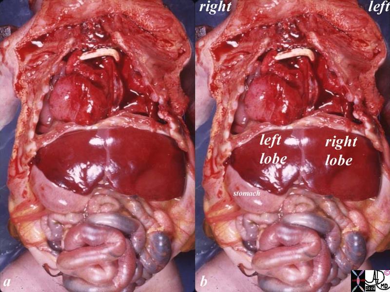
| 10850c01.8s post mortem baby with severe congenital anomalies including situs inversus totalis, dextroardia left sided liver right sided stomach severe congenital heart disease shunt possibly a Blalock Taussig large right ventricle grosspathology position malposition Courtesy Ashley DAvidoff MD copyright 2009 all rights reserved |