Copyright 2008
Ashley Davidoff MD
Definition
Acute aortic syndrome” (AAS) is a complication of atherosclerotic disease of the aorta that is characterized by a classical clinical presentation of sudden acute searing pain in the chest or back, usually in an hypertensive patient. Since the pathogenesis of this disease results in acute distension of the aortic wall its pain characteristics simulate acute aortic dissection. Hence the same dramatic pain characteristics that occur in dissection apply to acute aortic syndrome, characterized by a sudden acute, severe, shearng or lancinating, pain that reaches maximum intensity at onset. Similarly though as well, the symptoms may be minimal or absent.
The clinical features and pathology of the two entities can be quite different. Whereas dissection is usually in a younger age group, acute aortic syndrome is seen in the older atherosclerotic age group and is more commonly associated with hypertension. Classic aortic dissection is characterized by the presence of intimal tear that usually progresssses to the media and then rapidly dissects for quite a distance (sometimes the entire aorta) and will usually have a reentrance site. Intramural hematoma is characterised by the presence of severe atherosclerotic plaque, with acute hemorrhage into the aortic media which may remain as a hematoma in the media or may progress to a dissection. The dissection though is usually limited because the atherosclerois and inflammatory changes and fibrosis present in the wall prevent s an easy route for progression. The rentrance of the dissection in AAS is usually absent and hence thrombosis of the focal dissection results.
|
Pain Similar to Acute Dissection Searing Chest or Back Pain of Acute Aortic Syndrome |
| 49640c06 chest pain character shearing sharp lancinating acute aortic syndrome aortic dissection penetrating ulcer acute aortic hematoma ruptured aorta ruptured dissection CTscan Courtesy Ashley Davidoff MD |

The cause of AAS is an acute mural hematoma. This may result from a hematoma into a plaque, into a penetrating ulcer, or a hemattoma in a focal dissection. The space occupation and sudden distension associated with the disease process by one of these disorders results in the classical pain syndrome. The disorders are best diagnosed by using a combination of non-contrast CT combined with a contrast enhancing CT.
The hematoma in the wall may be complicated by a classicial dissection , where the tear in the wall extends along the longitudianl plain. Aortic rupture can occur as well.
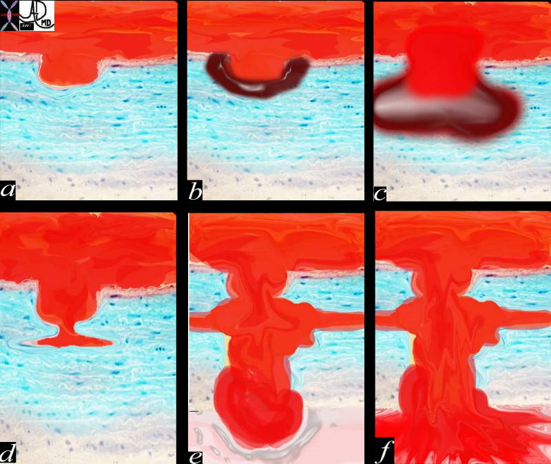
Pathogenesis of Penetrating Diseases of the Aorta and Acute Aortic Syndrome |
|
The lumen of the aorta with blood is demonsrated in red. The intima of the wall is the thin light pink layer just under the red lumenal layer. The media or elastic layer is in teal, while the outer layer is a light yellow color (a) This diagram illustrates the progression from a ulcerated plaque (a), to a mural hematoma(b). The ulcer in c has penetrated the into the media itself and is called a pentrating ulcer with a mural hematoma. (c). In d, a smal focal dissection with flowing blood is seen, and this can either thrombose or progress to a full dissection(e) or penetrate through the wall and be contained (e) or eventually rupture into the mediastinum (f)
42409c01.800 aorta artery atherosclerosis atheroma acute aortic syndrome a aortic ulcer b = acute mural hematoma c = acute mural hematoma large d = focal dissection e penetrating ulcer f rupture histology histopathology Davidoff art pathogenesis Courtesy Ashley Davidoff MD |
Treatment is based on the size position and nature of the lesion, as well as comorbid conditions, and thus would be either surgical or medical.
The pain is very similar to that of acute aortic dissection in that it is acute, severe, shearng or lancinating, but the clinical features and pathology of dissection is different. Whereas dissection is usually in a younger age group acute aortic syndrome is in the older atherosclerotic age group and is more commonly associated with hypertension. Classic aortic dissection is characterised by the presence of intimal tear that usually progresssses to the media and then rapoidly dissects for quite a distance (sometimes the entire aorta) and will usually have a reentrance site. Intramural haematoma is characterised by the presence of severe atherosclerotic plaque, with acute hemorrhage into the aortic media which may remain as a hematoma in the media or may progress to a dissection, but the dissection usually is limited beacuse the atherosclerois and inflammatory changes and fibrosis in general prevents easy route for progression. The rentrance of the dissection is usually absent and hence the focal dissection usually thrombosis.

Penetrating Ulcer |
| 48363 descending thoracic aorta fx aortic ulcer fx atherosclerosis atheroma fx penetrating ulcer CTscan Courtesy Ashley Davidoff MD |
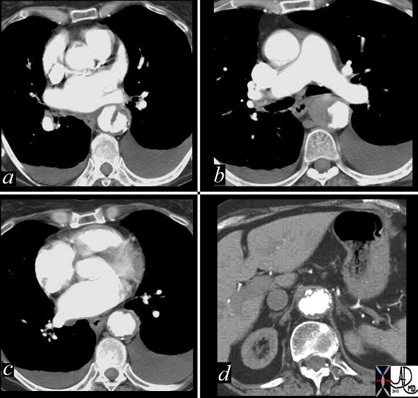
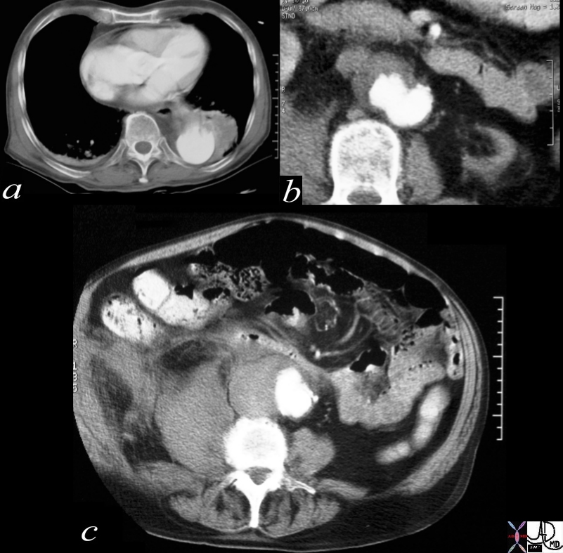
Penetrating Ulcer Mural Hematoma and Focal Dissection |
| This combination of images reflects the CTscan of a patient who presented with acute chest pain, and has findings reflecting a focal dissection of an atheromatous aorta. Note in (a) how thick the “intimal” flap is. Note also the mural dissection or hematoma with no flow within it. (b) Other parts of the descending aorta (c) and abdominal aorta (d) show only severe atheromatous disease. Courtesy Ashley Davidoff MD. 19416c code CVS aorta abdomen thorax dissection focal atherosclerosis |
Penetrating Ulcer with Rupture |
| 17529c01 artery descending thoracic aorta abdominal aorta dx rupture pseudoaneumysm ulcerating plaque mural hematoma ruptured through aortic wall hemorrhage hematoma retroperitoneum CTscan Courtesy Ashley DAvidoff MD Ashley Davidoff MD |
|
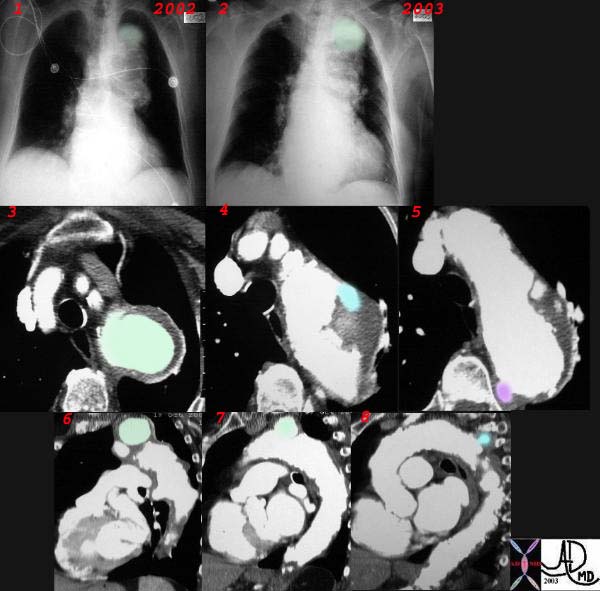
Penetrating Ulcer |
| This combination CXR and CT reveals an expanding aneurysm of the arch fromm 2002 to 3003. The CT shows three aneurysm in the arch of the aorta. The largest (green)seen in image 3,6, and 7 accounts for the finding in the left apex of the CXR, while a second pseudoaneurysm is seen on the lateral border of the knob (blue – 4,8) and a penetrating ulcer medially (purple 5) 32029c2 |