Right Ventricular Outflow Tract

The Common Vein

Copyright 2009


The Positional Relationship between the RVOT and the the Aortic Valve
The anatomic specimen (above) shows the right ventricular outflow tract (RVOT – blue dot) anterior and to the left of the aortic valve (AoV – red dot) which lies posterior and to the right.
The CT scan in axial projection through the same region shows the same relationship.
Ashley Davidoff MD


Normal Position of the Right Ventricular Outflow Tract (RVOT) in Relation to the Aortic Valve (AoV)
An axial black blood image of the through the atria and great vessels reveal the normal relative position of the RVOT in relation to the (AoV).
The RVOT lies anterior and leftward in relation to the aortic valve.
If we imagines d a clock then the RVOT is at about 1 o’clock
key words
heart cardiac aorta aortic valve infundibulum RVOT right ventricular outflow tract position relation MRIscan
Ashley Davidoff MD
07954b Wduo

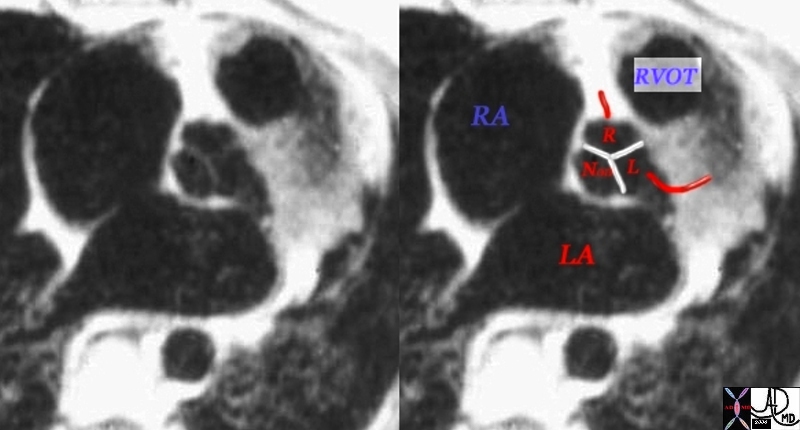
The inflow and outflow components of the RV are anatomically and embryologically distinct. The RVOT is tubular in shape and elevates the pulmonary valve above aortic valve. The chamber is thin walled, and twists around the aorta so that the pulmonary valve ends up to the left of the aortic valve. We are nearing the story of the “twist of the outflow tracts”


Normal Position and Relationship of the Aorta and Pulmonary Artery
This axial MRI view shows the RVOT anterior and leftward of the aortic valve, after it has twisted around the aorta. The thin musculature of the ovoid infundibulum is in red overlay in the second image.
Ashley Davidoff MD
07523e.81sL


Right Ventricular Enlargement and Hypertrophy
CT scan reconstructed in the sagittal plane shows right ventricular enlargement with hypertrophy of the trabeculated inflow portion and smooth walled outflow (RVOT) portion.  The RVOT points anteriorly.  The MPA also appears dilated.  The left atrium is enlarged and is compressing on a dilated esophagus.  The anterior leaflet of the mitral valve is in fibrous continuity with the aortic valve.
key words
infundibulum right ventricle RVOT right ventricular outflow tract aortic valve aortic sclerosis calcification enlarged left atrium heart normal conotruncal relationship cardiac
Courtesy Ashley Davidoff MD 2019
31149b.8s

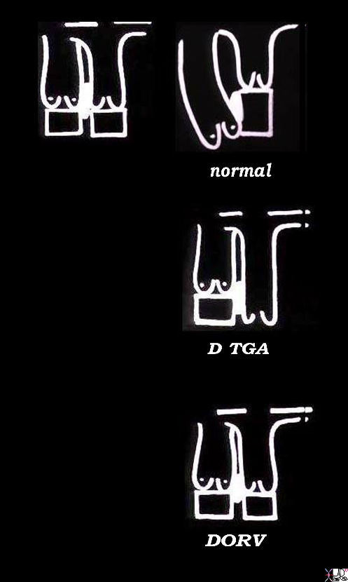
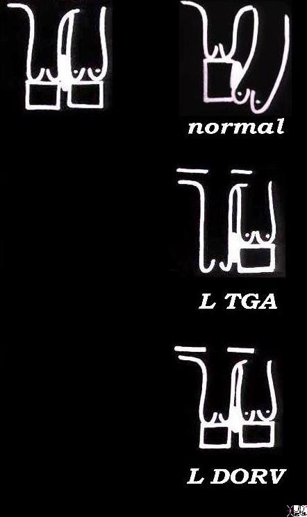
The “twist” in the story is long and complicated, with important implications in congenital heart disease – beyond the scope and need of this anatomy text. In the normal setting we make the following observations.

  1. The blue blooded chambers tend to be rightward, anterior, and slightly inferior.
  2. One of the main structural differences between the right and left sides is that the right ventricle has a separate and distinct outflow chamber – the RVOT or infundibulum.
  3. The presence and oblique leftward pointing orientation of this outflow chamber changes the relationship noted in #1, so that the pulmonary valve lies to the left and superior to its counterpart, the aortic valve.

During embryological development, the RVOT, MPA (Main Pulmonary Artery), and aorta twist around each other to result in the structural positioning and relationships described above. Under-twisting or over-twisting results in malpositioning of the great vessels in relation to their ventricles with consequent drastic hemodynamic results. Transposition of the great vessels is one such example.


 module76_07523%20trio%20550.jpg

Normal Position and Relationship of the Aorta and Pulmonary Artery

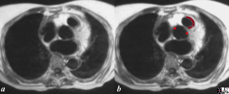
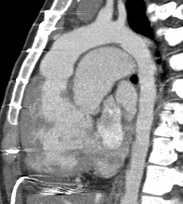
The “twist” story is graphically demonstrated in the following axial MRI images, cutting from inferior to superior as we follow the RV sinus, and RVOT around the aorta. Image 1 shows the triangular RV sinus anterior and to the right of the left ventricle. Image 2 is more superior and shows the RVOT making its way leftward and anterior to the aorta. Image 3 shows the final, distal, and leftward position of the RVOT just below the pulmonary valve. The next set of images with color overlay may be helpful.

Davidoff MD

module76_07523%20trio%20c%20W%20arrow%2002%20550.jpg

Normal Position and Relationship of the Aorta and Pulmonary Artery

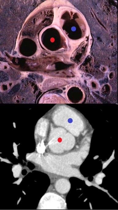
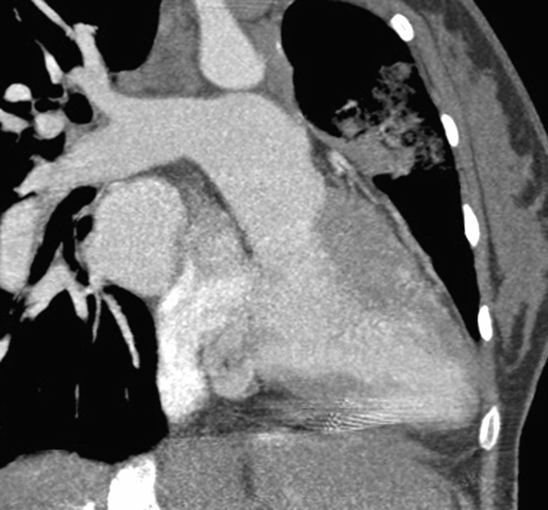
The “twist” of the story is now presented in color with Cupid’s arrow. Image 1 shows the triangular RV sinus anterior and to the right of the left ventricle, with arrow oriented to an 11 o’clock position. Image 2 is more superior and shows the RVOT making its way leftward and anterior to the aorta, with arrow oriented to an 12 or 1 o’clock position. Image 3 shows the final, distal, and leftward position of the RVOT just below the pulmonary valve with arrow oriented to a 1 or 2 o’clock position.

Davidoff MD (Images courtesy of Ashley Davidoff M.D.)


48321 CT of patient with an anterior aorta and a large posterior pulmonary artery TGA transposition heart aorta position 48320 48321 DTGA Courtesy Dr Hyun Woo Goo from Korea and Dr Laureen Sena

Diagram of the Embryological origins of Conotruncal Malformations
embryology bilateral conus DORV growth resorption normal mitral to aortic continuity transposition D transposition double outlet right ventricle
Ashley Davidoff art copyright 2019
06394c01L.800s
CT of patient with an anterior aorta and a large posterior pulmonary artery TGA transposition 48320 48321 DTGA
Courtesy Dr Hyun Woo Goo from Korea and Dr Laureen Sena

48320

 embryology bilateral conus DORV growth resorption normal mitral to aortic continuity transposition L transposition corrected transposition double outlet right ventricle Davidoff art copyright 2009 all rights reserved 06394c02Ls

< prev   next >