
IVC level sign
Shock
- Diagnosis
- clinical
- altered mental status,
- cool extremities and/or
- low urinary output.
- persistent hypotension
- systolic blood pressure < 80-90 mm Hg or
- mean arterial pressure 30 mm Hg lower than baseline
- reduction in cardiac index
- <1.8 L • min−1 • m−2 without support or
- <2.0 to 2.2 L • min−1 • m−2 with support
- echocardiography,
- deceleration time of early left ventricular filling has been shown to correlate inversely with pulmonary capillary wedge pressure,
- CT
- incidental radiographic findings
- dependent layering of contrast within the IVC and its tributaries
- creating a blood-contrast level.
- = IVC level sign
- incidental radiographic findings
- clinical
(top image)
Ashley Davidoff MD TheCommonVein.net
IVC LEVEL SIGN WITH NORMAL HEMODYNAMICS
An incidental Finding an a CTPA patient who was an outpatient who presented with elevated d dimer but was hemodynamically stable.
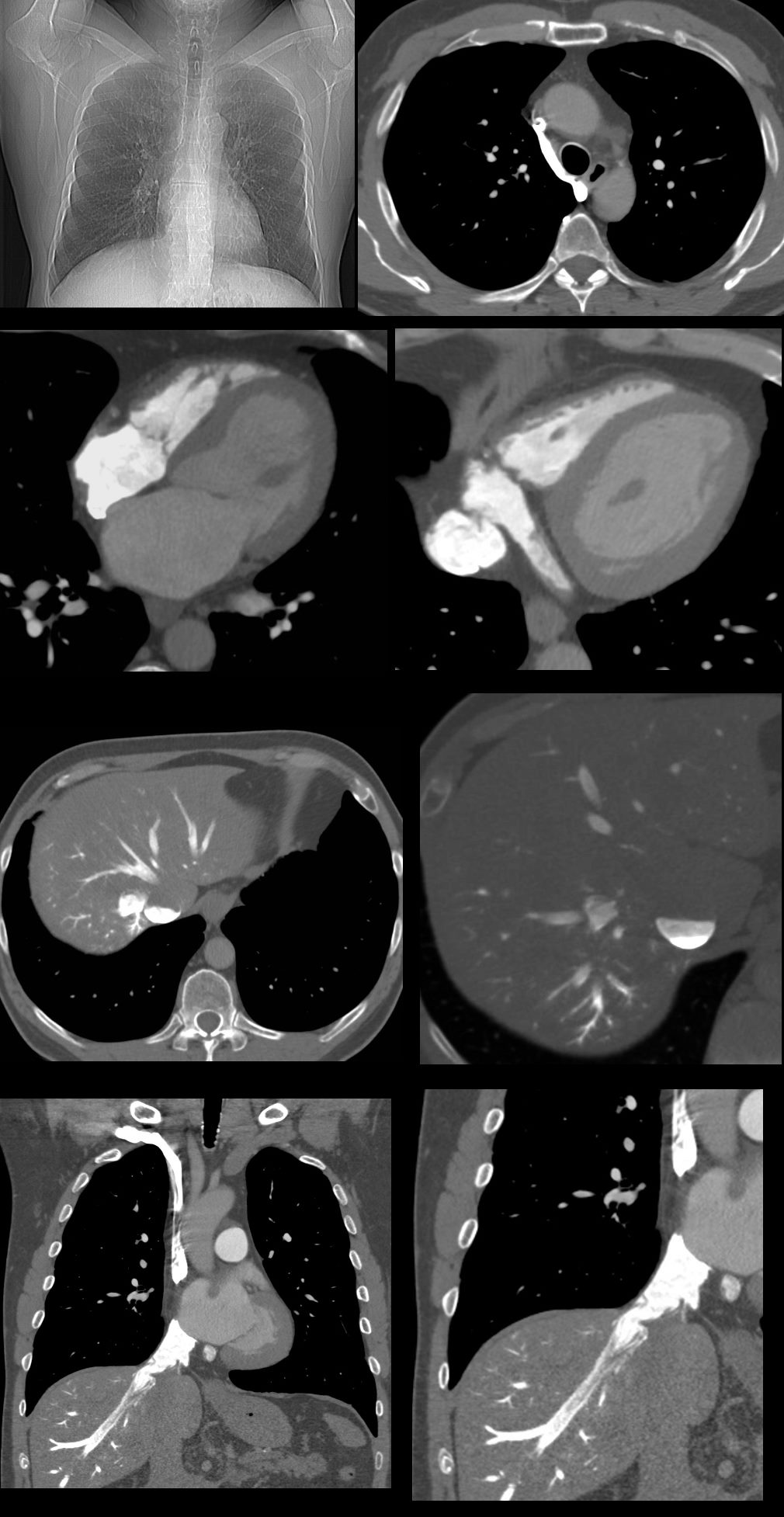
CT scan shows reflux into the azygous vein (top right), with normal to small right sided chambers, enlarged left atrium and left ventricle, bulging of the septum primum to the right indicating that left atrial pressures are higher than the right and enlarged IVC and coronary sinus.(second row)
There is significant stasis of systemic venous inflow into the right
atrium and right ventricle characterized by almost complete stasis of the
contrast column in the IVC and extension of the contrast into the smallest
hepatic veins in the dependent portion of the liver posteriorly. (3rd and 4th row) The blood contrast level in the IVC is called the “IVC level sign” (aka IVC niveau sign) Since this patient was completely well the explanation may relate to an unusually robust breath hold and [possible Valsalva maneuver during the study
Ashley Davidoff MD TheCommonVein.net See 82H
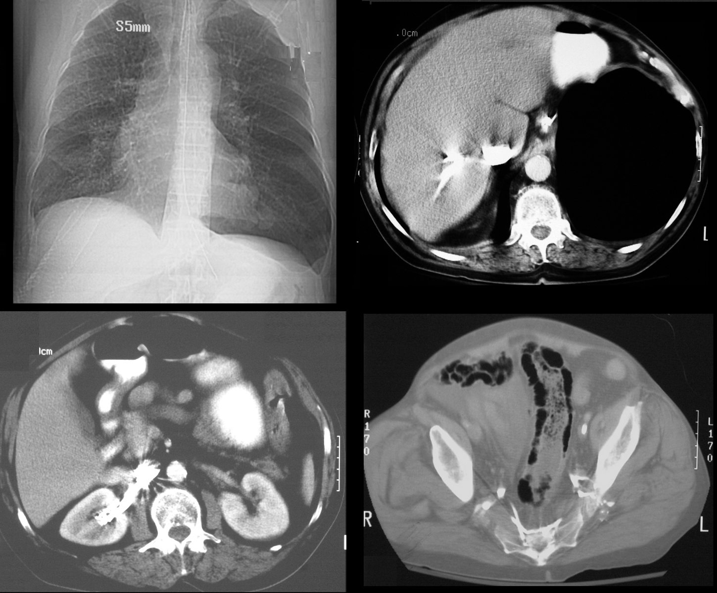
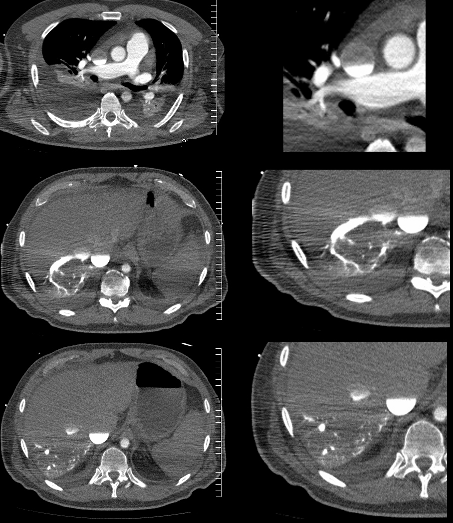
65 year old male s/p MVA presents in shock. Scout film (top left) shows left sided tension pneumothorax with rightward mediastinal shift. Axial CT through the liver (top right) shows expanded pneumothorax at the left lung base with reflux of contrast into the IVC.. Contrast also refluxes into the right renal vein (bottom left) and into the internal iliac veins (bottom right) Associated pneumatosis intestinalis in the sigmoid colon is present as well and likely secondary to the tension pneumothorax
Ashley Davidoff MD TheCommonVein.net
-
Links and References
-
Sullivan, I, W et al Dependent layering of venous refluxed contrast: A sign of critically low cardiac output Radiol Case Rep. 2019 Feb; 14(2): 230–234.
-
- 8. Tsai P.P., Chen J.H., Huang J.L., Shen W.C. Dependent pooling: a contrast-enhanced sign of cardiac arrest during CT. AJR Am J Roentgenol. 2002;178(5):1095–1099. [PubMed] [Google Scholar] [Ref list]
- 9. Ko S.F., Ng S.H., Chen M.C., Lee T.Y., Huang C.C., Wan Y.L. Sudden cardiac arrest during computed tomography examination: clinical findings and “dense abdominal veins” on computed tomography. J Cardiovasc Comput Tomogr. 2003;27(1):93–97. [PubMed] [Google Scholar] [Ref list]
- 10. Yeh B.M., Kurzman P., Foster E., Qayyum A., Joe B., Coakley F. Clinical relevance of retrograde inferior vena cava or hepatic vein opacification during contrast-enhanced CT. AJR Am J Roentgenol. 2004;183(5):1227–1232. [PubMed] [Google Scholar] [Ref list]
- 11. Roth C., Sneider M., Bogot N., Todd M., Cronin P. Dependent venous contrast pooling and layering: a sign of imminent cardiogenic shock. Am J Roentgenol. 2006;186(4):1116–1119. [PubMed] [Google Scholar] [Ref list]
- 12. Sueyoshi E., Imamura T., Sakamoto I., Uetani M., Matsuoka Y. Contrast-fluid level in the inferior vena cava (IVC niveau sign) in patients with acute type A aortic dissection: computed tomography findings during acute cardiac tamponade. Jpn J Radiol. 2010;28(4):278–282. [PubMed] [Google Scholar] [Ref list]
- Bagheri S.M., Taheri M.S., Pourghorban R., Shabani M. Computed tomographic imaging features of sudden cardiac arrest and impending cardiogenic shock. J Comput Assist Tomogr. 2012;36(3):291–294. [PubMed] [Google Scholar] [Ref list]
- 14. Wong H.Y., Lee K.H. The IVC contrast level sign. Abdom Radiol. 2017;42(12):2962–2963. [PubMed] [Google Scholar] [Ref list]
- 15. Shiotani S., Kohno M., Ohashi N., Yamazaki K., Itai Y. Postmortem intravascular high-density fluid level (hypostasis): CT findings. J Comput Assist Tomogr. 2002;26(6):892–893. [PubMed] [Google Scholar] [Ref list]
-
TCV