
- Buzz
- Myxoma = myxoid, hypocellular
- poorly enhancing
- water/jelly (low density t2)
- floppy – prolapse through the valve
- trauma
- hemorhage (T1 and T2 variability)
- calcification (T2 dark)
- hemorhage (T1 and T2 variability)
- trauma
- embolize
- floppy – prolapse through the valve
- Rare but most common benign tumor of the heart (50%)
- Myxoma = myxoid, hypocellular
- Atrial Myxoma
- is a benign neoplasm
- characterised by
-
- Structural Features
- Left Atrium (> 80%), (fossa ovale)
- if in right side or LV multicentric more common
- Attached to the septum
- Often prudes through the A-V valve
- Sessile or pedunculated
- repeated trauma they hemorrhage and therefore calcify
- Left Atrium (> 80%), (fossa ovale)
- Functional Features
- mechanical obstruction of the LA
- embolisation
- Structural Features
-
- Etiology
- 90% arise sporadically
- 10% inherited (eg Carney Syndrome)
- Pathogenesis
- derived from multipotent mesenchymal cells.
- Resulting in
- Pathology
-
ATRIAL MYXOMA
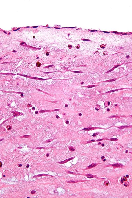
High magnification micrograph of an atrial myxoma. H&E stain. Atrial myxoma is the most common primary tumour of the heart. It is benign. Features: Hypocellular myxoid polypoid mass with cells that are: Perivascular arrangement, Finely vacuolated eosinophilic cytoplasm, Polygonal/elongated cell shape, Mononuclear or multinucleated. May contain macrophages, other inflammatory cells. Often covered with endothelium.
Courtesy Wikipedia
-
- Pathology
- Diagnosis
- Imaging
- Echo
- See Echo (wiki)
- See Echo (wiki)
- CT
- Echo
-
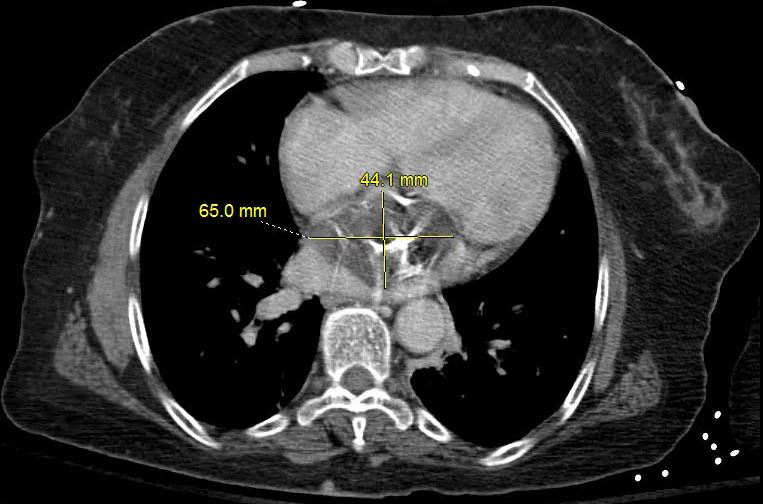
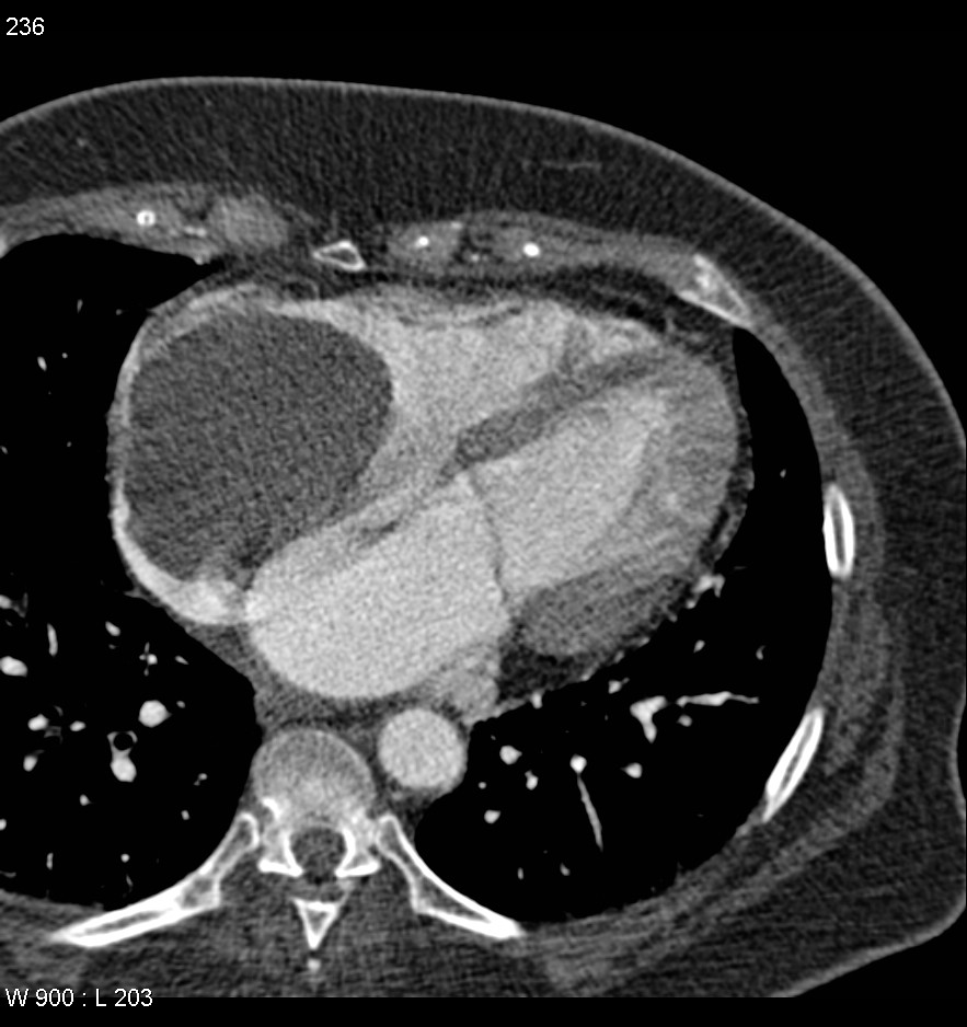
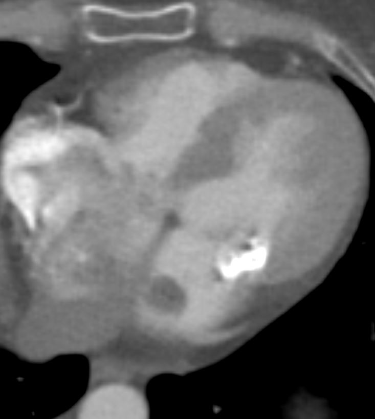
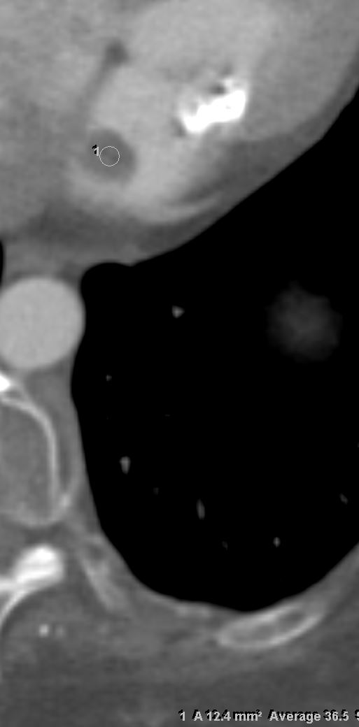
- 66-year-old female presents with chest pain
-
- The frontal CXR shows left atrial enlargement characterized by elevated L main stem bronchus, and in retrospect some calcifications overlying the left atrium on the lateral
-
- The scout film prior to the CT confirms the elevated left main stem bronchus, but the dominant finding is a calcified left atrial mass that is attached to the atrial septum. It measures 6.5 X 4.4 X 4.6cms and shows mild enhancement.
-
- The other chambers are normal in size
-
- The mass was resected and pathology findings were consistent with an atrial myxoma
- Ashley Davidoff MD

CALCIFIED LEFT ATRIAL MYXOMA
66-year-old female presents with chest pain
The frontal CXR shows left atrial enlargement characterized by elevated L main stem bronchus, and in retrospect some calcifications overlying the left atrium on the lateral
The scout film prior to the CT confirms the elevated left main stem bronchus, but the dominant finding is a calcified left atrial mass that is attached to the atrial septum. It measures 6.5 X 4.4 X 4.6cms and shows mild enhancement.
The other chambers are normal in size
The mass was resected and pathology findings were consistent with an atrial myxoma
Ashley Davidoff MD
Ashley Davidof MD TheCommonVein.net
Ashley Davidof MD TheCommonVein.net
MRI
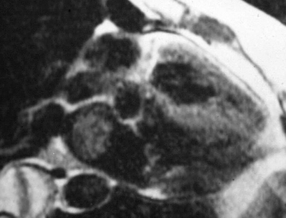
ATRIAL MYXOMA
58-year-old female presents with cough
The frontal CXR shows probable left atrial enlargement characterized by elevated L main stem bronchus (b black arrow), double density (red arrow, b) and a straight heart border caused mostly by a large PA segment (blue arrow b) and evidence of redistribution (a). The lateral confirms left atrial enlargement with posterior displacement and elevation of the left main stem bronchus (d, black arrow). The left ventricle is not enlarged. These findings suggest mitral stenosis.
Black blood sequence shows a heterogeneous mass in the left atrium (LA) attached to the septum with signal brighter than the myocardium and less than fat. These findings are characteristic of an atrial myxoma. The mass was removed and pathology confirmed an atrial myxoma
Ashley Davidoff MD
T2 – brighter than the myocardium less than fat
mobile
heterogenous mass
LGE
mass is heterogeneous,
some enhancement
References and Links
Hoey E., Shahid M, Ganeshan A. et al., MRI assessment of cardiac tumours: part 1, multiparametric imaging protocols and spectrum of appearances of histologically benign lesions, Quant Imaging Med Surg, 4 (6), 12/2014; 478-488