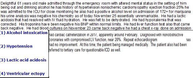
HISTORY AND CLINICAL
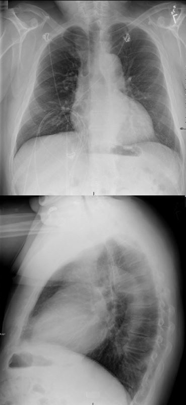
CXR shows LV configuration on the frontal and LVE on the lateral
Ashley Davidoff MD
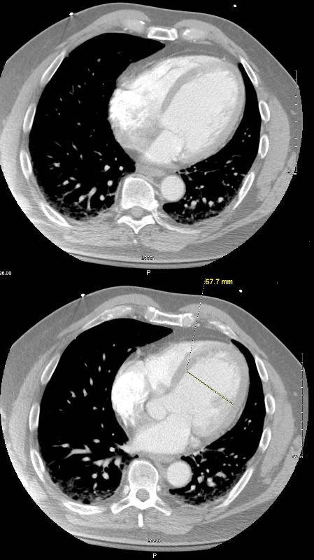
Axial CT through the 4 chambers shows isolated LV dilatation in early diastole with the cavity measuring 6.8cms.
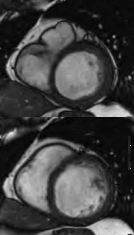
MR in axial plane shows a dilated LV and normal sized LA, RA, and RV. EF was between 10% and 20%
Ashley Davidoff MD
61 male, alcoholic with congestive cardiomyopathy
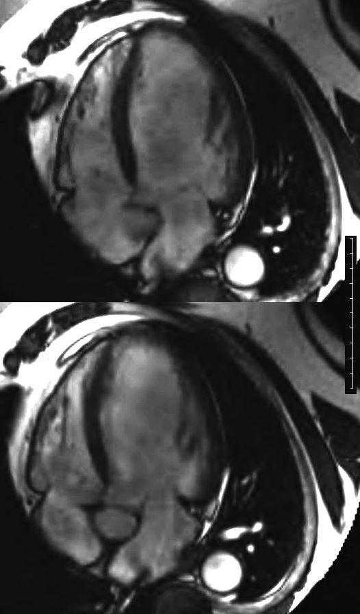
MRI in short axis through the body of the LV in systole (above) and diastole (below)shows a dilated LV without much change in the dimensions of the LV cavity. EF was between 10% and 20%
Ashley Davidoff MD
CONGESTIVE CARDIOMYOPATHY
61 male, with alcoholic congestive cardiomyopathy.
MRI of the heart in short axis, shows the LV in systole (above) with the mitral valve (MV) closed and in diastole (below) with the open mitral valve.
Ashley Davidoff MD
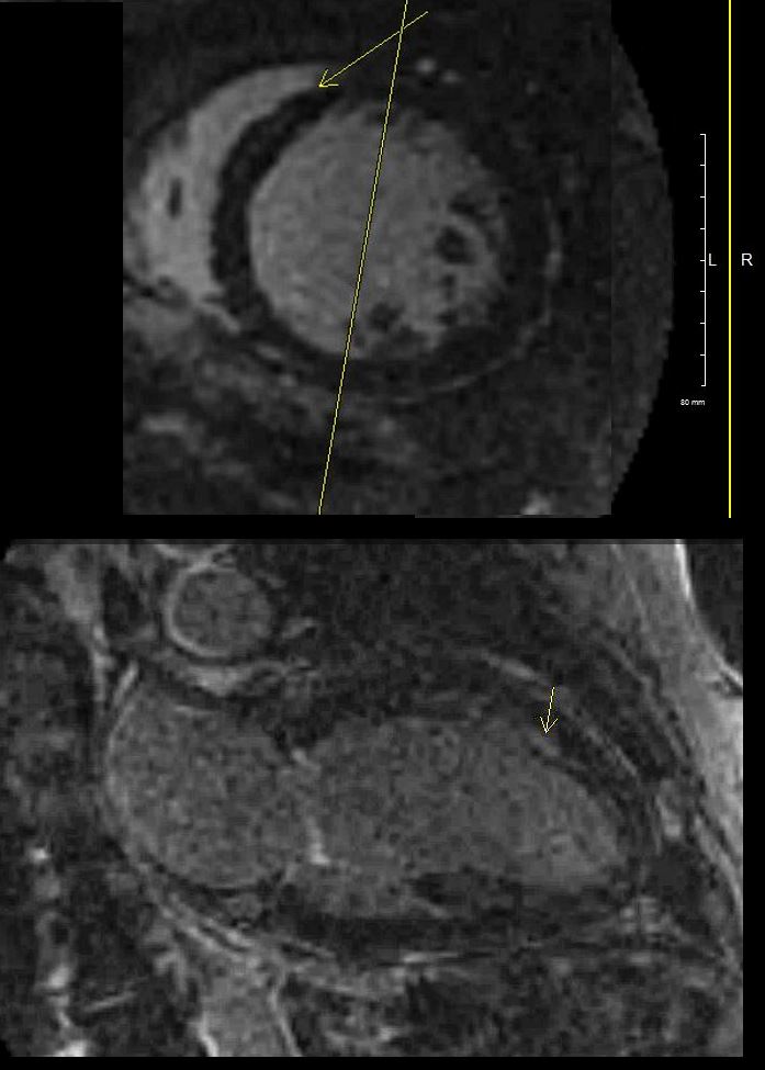
LGE

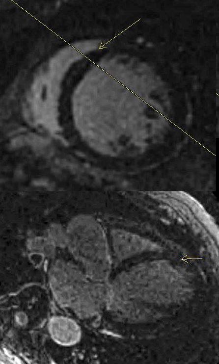
Short axis LGE sequence shows linear mid myocardial LGE in the ventricular septum
Ashley Davidoff MD
CONGESTIVE CARDIOMYOPATHY
61 male, alcoholic, with congestive cardiomyopathy
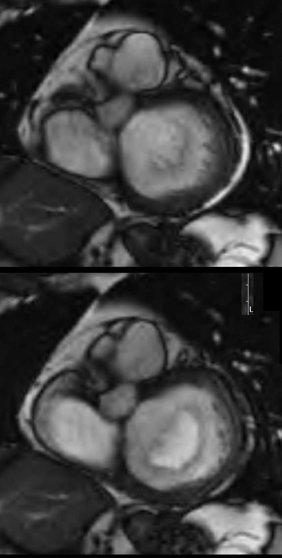
4 chamber LGE sequence shows linear mid myocardial LGE in the ventricular septum
Ashley Davidoff MD
2 Chamber LGE sequence shows linear mid myocardial LGE in the ventricular septum
Ashley Davidoff MD