
Mitral Annular Calcification (MAC)
Copyright 2008
- Degenerative process associated with
- aging,
- cardiovascular risk factors.
- Degeneration from increased mitral valve stress
- Aortic Stenosis
- Hypertension
- Hypertrophic Cardiomyopathy
- Also seen in
- chronic kidney disease, (Ca PO4 disorder)
- following radiation therapy.
- Associated with
- arrhythmias and heart blocks
- MS and MR are rare
Definition
Mitral annular calcification is a degenerative disorder of the mitral annulus that occurs in 6% of elderly patients. Although it is part of the aging process it is a marker for other atherosclerotic diseases, and is commonly associated with aortic sclerosis which also occurs in similar patient populations. It has been suggested that MAC and aortic stenosis have a close link (Movahed)
Structurally at a histological level fibrillar degeneration of the collagen appears to be the precipitating event followed by dystrophic calcification. The calcification spares the portion of the annulus that is attached to the anterior leaflet of the mitral valve so that it is shaped like a backward “C” in the A-P projection and even in the lateral projection.
Functionally it often has no clinical bearing, but when severe it may cause mitral regurgitation, stenosis, or even extend into the conduction system where heart block may result.
It is best diagnosed by plain film but usually milder forms are not visualized. Echocardiography and CTscanning easily diagnose this condition.
Since it is a marker of cardiovascular disease treatment is directed at the associated conditions rather than at MAC.
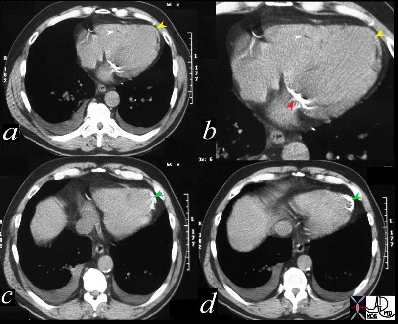
56 year old male with history of coronary artery disease. Axial CT through the heart shows apical curvilinear fat (yellow arrowheads, ( a and b) associated with apical myocardial dystrophic calcification (green arrowheads c and d) both indicating prior apical MI. In addition there mitral annular calcification (red arrowhead, b) and multifocal fatty deposits in the RV (white arrowheads, a and b) usually depicting age related degenerative changes. The calcification in the annulus is premature and unusual for this 56 year old male patient. Note the small bilateral pleural effusion The association of atherosclerosis and MAC are well known. Premature MAC should therefore be a warning for premature heart disease.
Ashley Davidoff MD
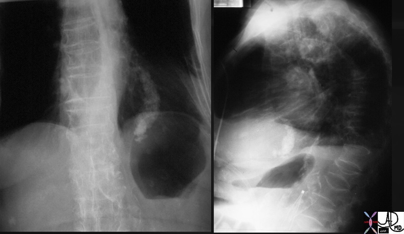
MAC in an Elderly female |
| Note the incomplete ring of calcification. Calcification is absent in the anterior leaflet of the mitral valve
32744c01 heart mitral valve mitral annulus mitral annular calcification MAC aortic calcifications osteopenia bone thoracic spine kyphosis compression fracture CXR plain film of the chest IVC filter Davidoff MD |
MAC Extending Down the Interventricular Septum |
| 34130 heart mitral valve mitral annulus mitral annular calcification MAC progression down the interventricular septum conduction defects nerve involvement CTscan Davidoff MD |
Mitral Annulus and Aortic Annulus Calcification
MAC and IHD
56 year old male with history of coronary artery disease. Axial CT through the heart shows apical curvilinear fat (yellow arrowheads, ( a and b) associated with apical myocardial dystrophic calcification (green arrowheads c and d) both indicating prior apical MI. In addition there mitral annular calcification (red arrowhead, b) and multifocal fatty deposits in the RV (white arrowheads, a and b) usually depicting age related degenerative changes. The calcification in the annulus is premature and unusual for this 56 year old male patient. Note the small bilateral pleural effusion The association of atherosclerosis and MAC are well known. Premature MAC should therefore be a warning for premature heart disease.
Ashley Davidoff MD
Complications
Heart Block, MR and MS
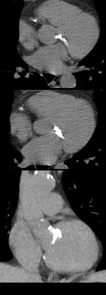
82-year-old female with end stage renal failure, diabetes mellitus, CAD, s/p CABG x2 and AVR, severe mitral annular calcification, extending to the septum, s/p pacemaker, with associated mild mitral stenosis and significant mitral regurgitation. Severe aortic stenosis warranted AVR.
Axial CT through the mitral annulus shows severe mitral annular calcification extending into the ventricular cavity and the ventricular septum.
Ashley Davidoff MD
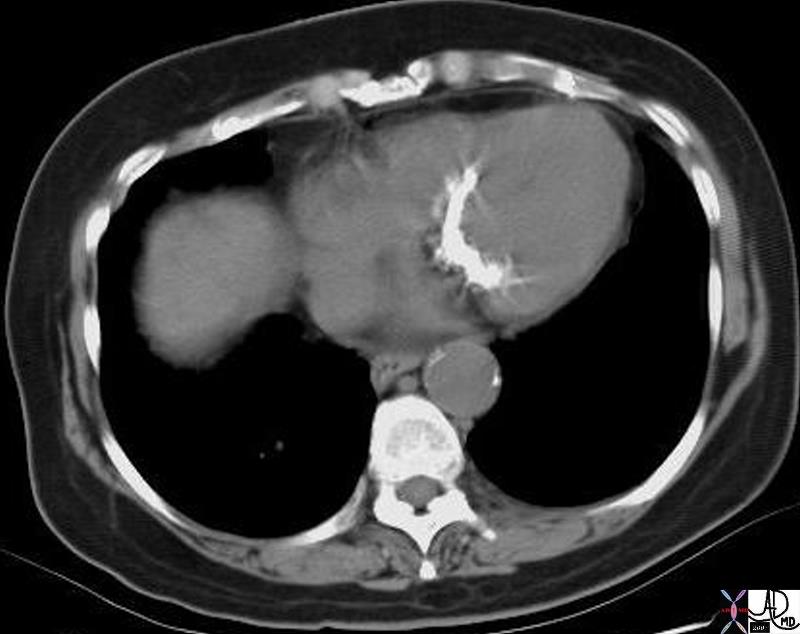
73 year old female with question of mass in the region of the mitral annulus consistent with a large collection of mitral annular calcification
Ashley Davidoff MD

73 year old female with question of mass in the region of the mitral annulus consistent with a large collection of mitral annular calcification
Ashley Davidoff MD
73 year old female with question of mass in the region of the mitral annulus consistent with a large collection of mitral annular calcification
Ashley Davidoff MD
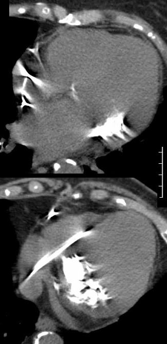
MAC and Caseous Necrosis

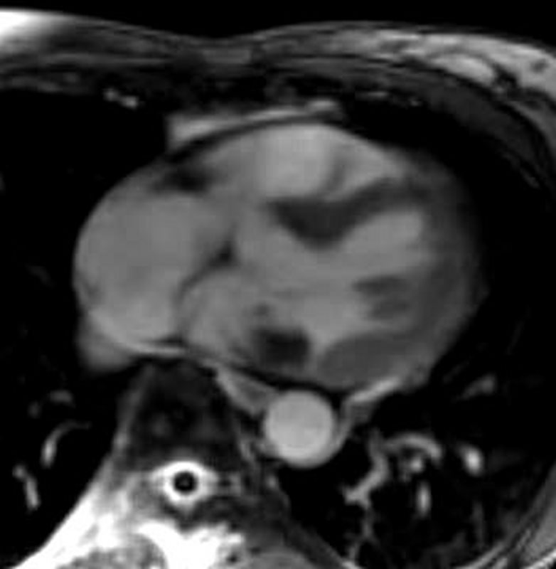
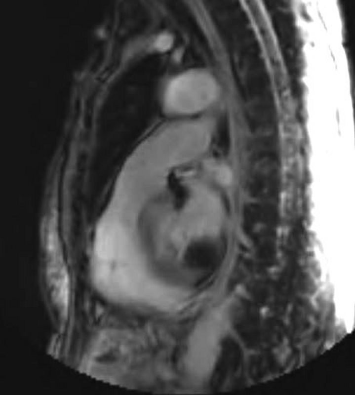
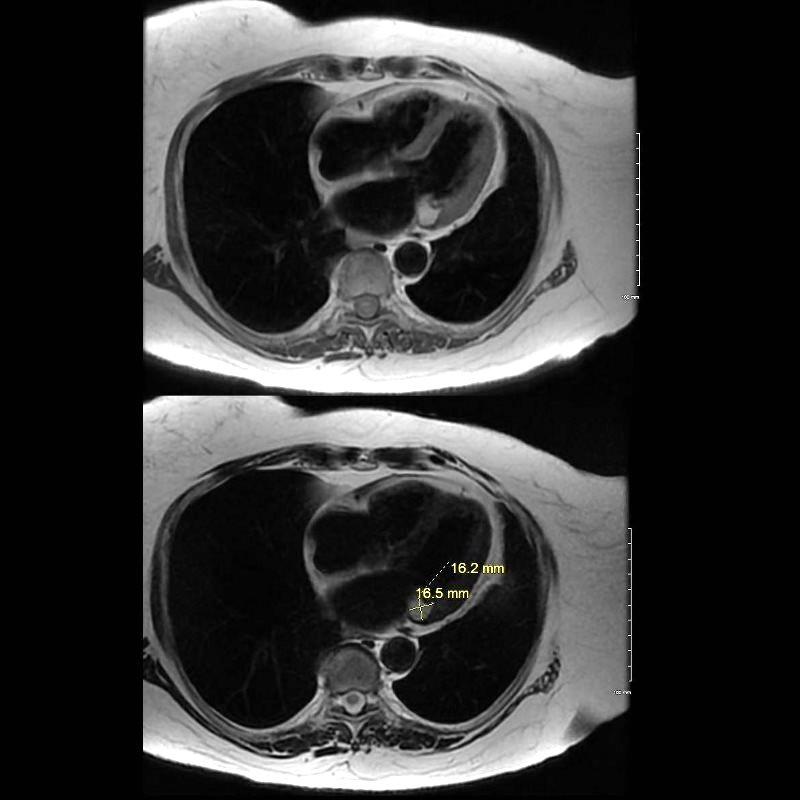
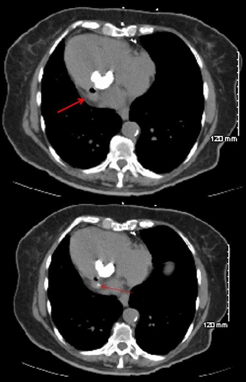
69 year old female with MAC who presents with an echo finding of a mass on the mitral valve thought to represent a myxoma. CT confirmed a low-density mass measuring in the soft tissue range associated with mitral annular calcification. MRI showed a mass in the region of the posterior leaflet of the MV with T2 hyperintensity suggesting aqueous component and T1 hyperintensity suggesting increased protein content. There was no enhancement of the lesion. Peripheral T1 and T2 low intensity reflects calcification. A diagnosis of caseous necrosis of MAC is most likely
Ashley Davidoff MD
. MRI showed a mass in the region of the posterior leaflet of the MV with T2 hyperintensity suggesting aqueous component and T1 hyperintensity suggesting increased protein content. There was no enhancement of the lesion. Peripheral T1 and T2 low intensity reflects calcification. A diagnosis of caseous necrosis of MAC is most likely
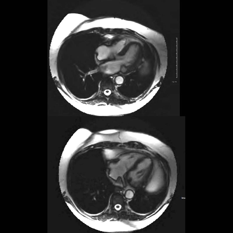
MRI showed a mass in the region of the posterior leaflet of the MV with T2 hyperintensity suggesting aqueous component (upper image) and focal T1 low intensity nodule on the posterior leaflet reflects the MAC There was no enhancement of the lesion. A diagnosis of caseous necrosis of MAC is most likely
MAC Dextrocardia and Bacterial Endocarditis
78 year old female with dextrocardia, presents 2 weeks after laparoscopic surgery with a fever. Prior CT showed dextrocardia with MAC. Repeat CT at the time of the fever, shows an air fluid on the lateral aspect of the MAC with disrupted calcification. Blood cultures were positive and a diagnosis of anaerobic gas forming endocarditis was made. She subsequently required MVR
Ashley Davidoff MD
Links and References
Abramovitz Y et al Mitral Annulus Calcification. J Am Coll Cardiol 2015;66:1934-1941. (excellent review)
Allison M, et al Mitral and Aortic Annular Calcification Are Highly Associated With Systemic Calcified Atherosclerosis Circulation Vol. 113, No. 6