
-
The Heart
- Left Heart
- PV
- LA
- LV
- Right Heart
- RA
- SVC
- Azygous vein
- IVC
- RV
- PA
Left Heart
PV
LA

74-year-old female presents in CHF and an echo showing reduced EF (35%) and non compaction.
Initial CXR shows findings consistent with interstitial edema, (redistribution, fuzzy borders of the vessels and descending RPA) Kerley B lines, and left atrial enlargement.
Prior to implantation of a dual lead pacemaker she had a gated cardiac CT to define the venous anatomy.
The scout film shows an enlarged left atrium and suggestion of LV enlargement.
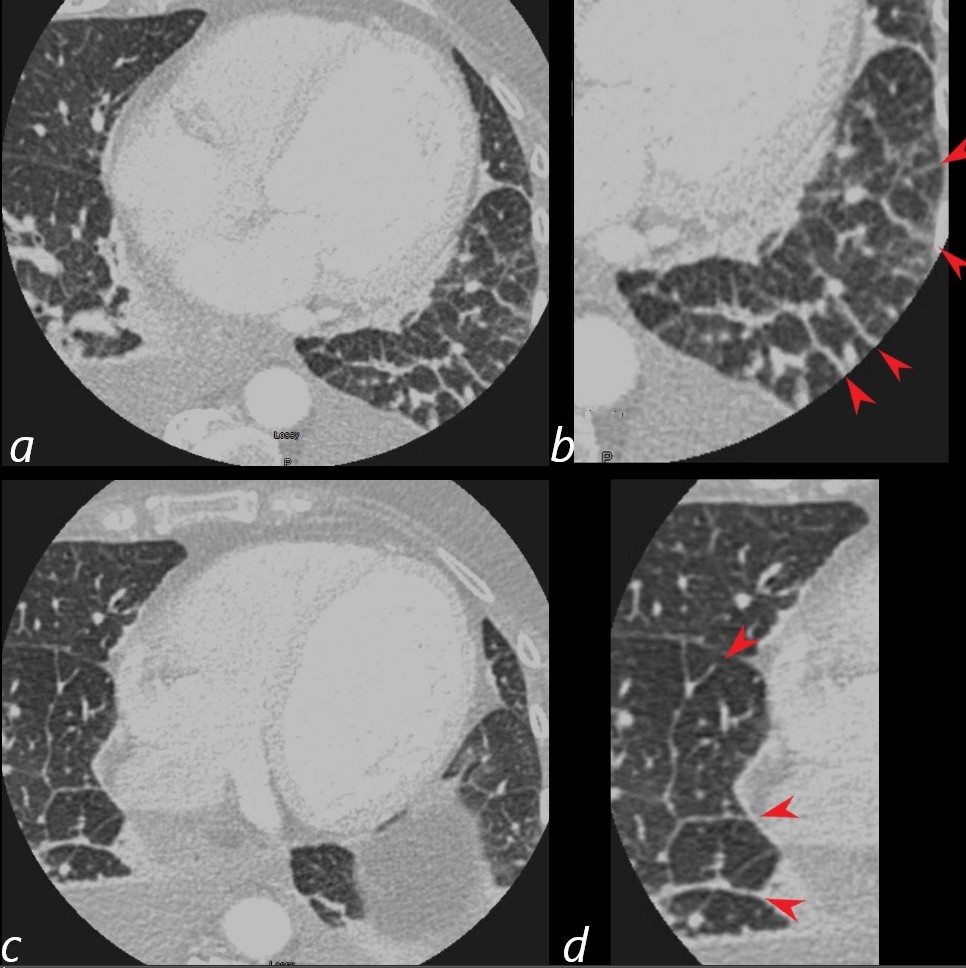
Lung windows confirmed the presence of prominent interlobular septa and LAE.
Axial soft tissue windows shows LAE with A_P dimension of 4.7cms (upper limits of normal is 4cms)
Ashley Davidoff MD
LV
NON-COMPACTION AND CHF
74-year-old female presents in CHF and an echo showing reduced EF (35%) and non compaction.
Initial CXR shows findings consistent with interstitial edema, (redistribution, fuzzy borders of the vessels and descending RPA) Kerley B lines, and left atrial enlargement.
Prior to implantation of a dual lead pacemaker she had a gated cardiac CT to define the venous anatomy.
The scout film shows an enlarged left atrium and suggestion of LV enlargement.
Lung windows confirmed the presence of prominent interlobular septa and LAE.
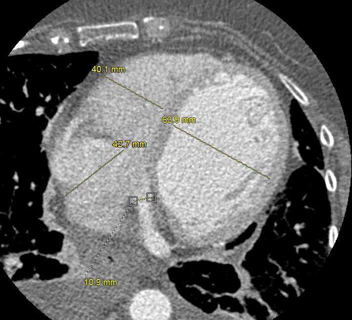
Axial soft tissue windows confirmed a diagnosis of non-compaction with non compaction thickness (NC) of 15mm and free wall thickness (C) of 6mm resulting in an abnormal NC:C ratio of 2.5 (upper limits normal NC:C ratio = 2.3)
Volume measurements showed an end diastolic volume of 217 mls, an end systolic volume of 159ccs, a stroke volume of 58ccs with a resulting ejection fraction of 26%.
Ashley Davidoff MD

74-year-old female presents in CHF and an echo showing reduced EF (35%) and non compaction.
Volume measurements based on the gated cardiac CT showed an end diastolic volume of 217 mls, an end systolic volume of 159ccs, a stroke volume of 58ccs with a resulting ejection fraction of 26%.
Ashley Davidoff MD
Lungs
74-year-old female presents in CHF and an echo showing reduced EF (35%) and non compaction.
Initial CXR shows findings consistent with interstitial edema, (redistribution, fuzzy borders of the vessels and descending RPA) Kerley B lines, and left atrial enlargement.
Prior to implantation of a dual lead pacemaker she had a gated cardiac CT to define the venous anatomy.
The scout film shows an enlarged left atrium and suggestion of LV enlargement.
Lung windows confirmed the presence of prominent interlobular septa and LAE with bilateral complex effusions.
Ashley Davidoff MD
Crazy Paving – CHF A Rare Cause
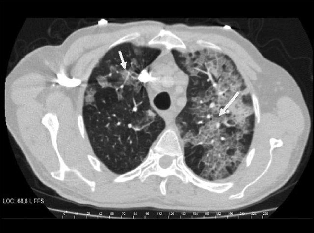
44-year-old man with dyspnea and cough diagnosed as having congestive heart failure. CT of the thorax shows diffuse, multifocal, patchy, ground-glass opacities (arrow).
Şentürk A, et al A Rare Cause of Crazy-Paving and Mediastinal Lymphadenopathy: Congestive Heart Failure Journal of Clinical Imaging Science 3(1):30 July 2013
Right Heart
RA t\
Tricuspid Regurgitation
SVC
Azygos vein
IVC
RV
PA
References and Links