
Levocardia Mesocardia Dextrocardia and Dextroposition
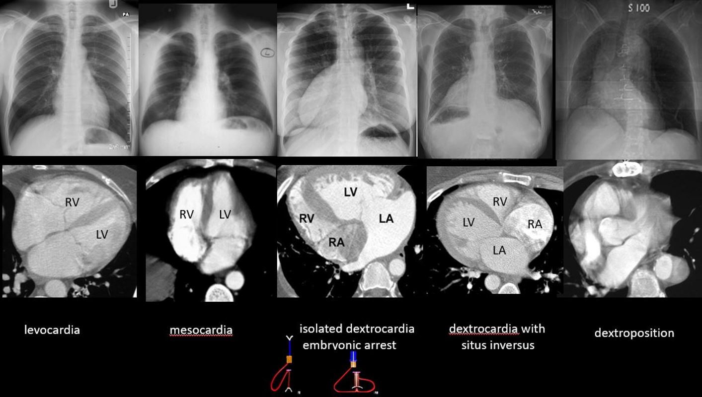
The collage reveals the variation in the position of the LV with the CXR ‘s above and the correlate CT below
The first row shows the normal appearance of the apex of the heart in the left chest reflecting the position of the LV. The correlate image below shows the normal position of the LV and its relation to the right ventricle (RV). Note the stomach bubble is on the left indicating situs solitus
The second column reflects mesocardia. On the CXR above the heart is almost midline in position and the CT image below shows the LV apex pointing forward and the chambers lie side by side. Note the stomach bubble is on the left indicating situs solitus
The 3rd column reflects isolated dextrocardia caused by an arrest of the normal looping of the heart. The RV forms first and normally loops to the right. The LV is a daughter of the RV and eventually grows to create the apex of the heart to the left. In this entity there is an arrest of that latter process and the RV assumes the responsibility to form the apex. Note the stomach bubble is on the left indicating situs solitus.
This entity is commonly associated with severe congenital heart abnormalities such as pulmonary hypoplasia.
In the next column, the apex of the heart is also pointing to the right and the stomach bubble is on the right as well. In this instance there is situs inversus totalis with the viscera, atria and ventricles being mirror images of the normal. Although congenital abnormalities do occur in this entity, they are less frequent than the entity of dextrocardia with situs solitus (“embryonic arrest” form)
The last column is an example of dextroposition of the heart because of reduction in volume of the right chest following surgical removal of parts of the right lung and the heart and left lung fill up that space
Ashley Davidoff MD
The Right Upper Cut
- Right Sided Chambers
- Inferior and Anterior
- Left side Chambers
- Superior and Posterior

Right Sided Structures Right Sided, Anterior and Inferior, Except the Pulmonary Artery which is Leftward and Superior
Cross Sectional Axial
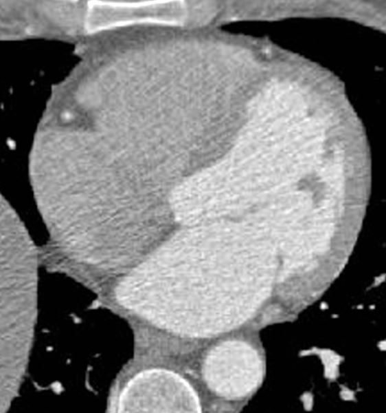
Axial images through the 4 chambers at the level of the A-V valves during diastole (mitral valve open) enables an approximate volume evaluation of the chambers. The atria are approximately the same volumes, and are about 1/3 the volume of the ventricles. The right ventricle (RV) is about 2/3 the volume of the left ventricle (LV)
Ashley Davidoff MD
Ashley Davidoff MD