
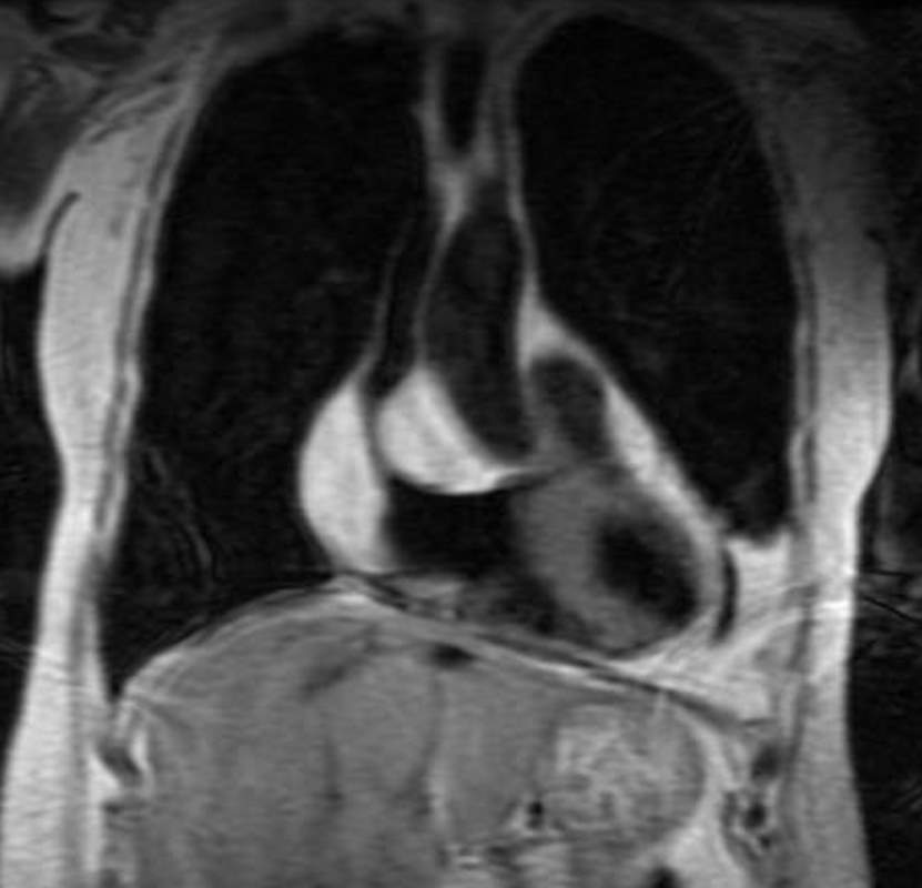
This image of a coronally acquired T1 weighted MRI image of the heart shows a high intensity mass surrounding the SVC and the entrance right atrium (RA) with narrowing of the SVC. There were no symptoms of SVC syndrome in this patient with known COPD. Note that the mass has the intensity of subcutaneous fat. An atrial lipoma is the most likely diagnosis.
Courtesy Jorge Medina
38449c
KEY WORDS
Cardiac, heart, vein, SVC , RA, mass, fat, lipoma, tumor, neoplasm, benign, imaging, radiology, MRI
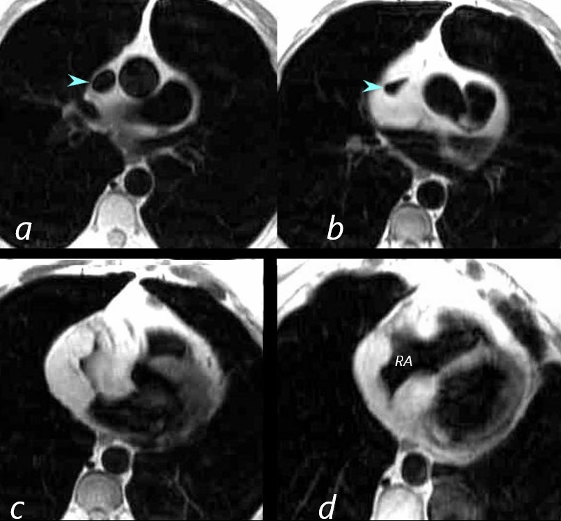
This series of axial T1 weighted MRI images of the heart show a high intensity mass surrounding the SVC (blue arrowhead) and the entrance to the right atrium (RA). There were no symptoms of SVC syndrome in this patient with known COPD. Note that the mass has the intensity of subcutaneous fat. An atrial lipoma is the most likely diagnosis.
Courtesy Jorge Medina
38449c
KEY WORDS
Cardiac, heart, vein, SVC , RA, mass, fat, lipoma, tumor, neoplasm, benign, imaging, radiology, MRI