The Common Vein
Copyright 2009
Definition
Cardiomyopathy is a general term reflecting cardiac muscle disease. It may be primary in which case the cause is unknown or secondary to an identifiable agent. The most common of these agents include ischemia, viral infection (esp. Coxsackie) and alcohol. Other common associated diseases include hypertension, valvular heart disease and congenital heart diseases. Less common causes include Adriamycin, amyloidosis endocardial fibroelastosis (EFE), and Pompe (glycogen storage) disease.The cardiomyopathies are classified according to anatomical and functional manifestations. The three main types of disease include dilated, hypertrophic, and restrictive cardiomyopathy. Dilated cardiomyopathy is the most common.The cardiomyopathies usually result in a combination of left and right ventricular dysfunction, with dyspnea and orthopnea reflecting left sided disease and with peripheral edema and hepatomegaly reflecting right sided disease.The diagnosis of cardiomyopathy is best made using echocardiography, where the distinction between dilated, hypertrophiic and restrictive disease can be made.Treatment depends on the type of cardiomyopathy, but usually involves medical management of heart failure, and if a surgical cause can be identified then appropriate management instituted.
|
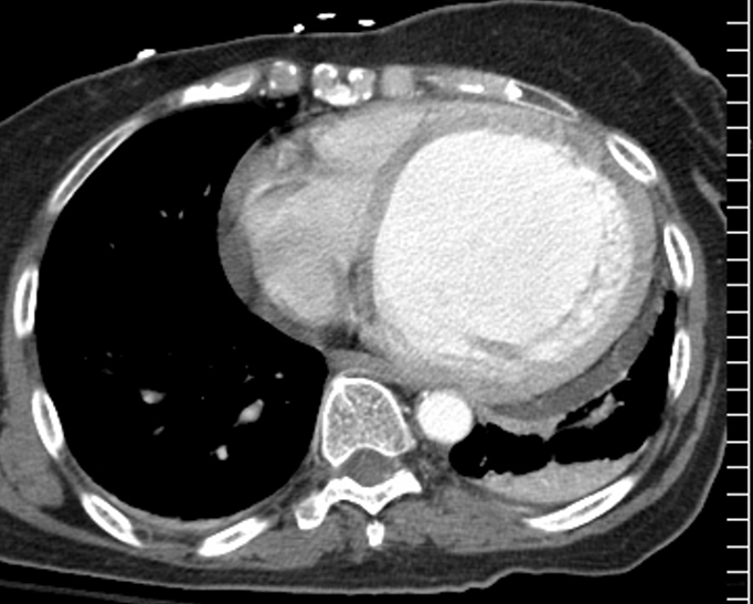
Dilated Cardiomyopathy |
| 31675b01 heart cardiac LV IVS interventricular septum left ventricle LVE left ventricular enlargement left ventricular dilatation cardiomyopathy size anemia thin septum CTscan Courtesy Ashley Davidoff MD |
|
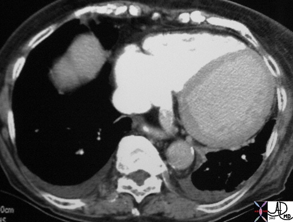
Dilated Cardiomyopathy |
| The chest CT is taken through the heart with a dense contrast phase in the right sided structures with very little contrast in the left. This is not normal and suggests pump failure. What do you think of the left ventricle? This is a globally dilated LV with a cardiomyopathy. Note the contrast in the coronary sinus which is also slightly enlarged indicating elevated right sided pressures. Courtesy Ashley Davidoff MD 26321 code LV dilated cardiomyopathy hypokinesis cardiac imaging radiology CTscan |
|
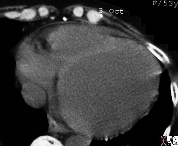
Dilated Cardiomyopathy |
| 75517 elderly female heart cardiac paillary muscle left ventricle fx dilated LV shape change size small pericardial effusion dx congestive cardiomyopathy fx atelectasis CTscan Courtesy Ashley Davidoff MD copyright TCV 2008 |
|
Dilated Cardiomyopathy |
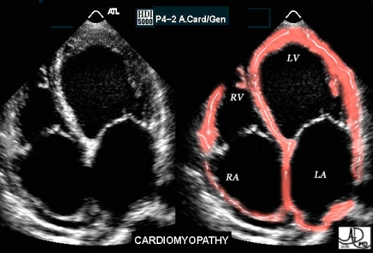
| This image is a real time echocardiogram, projecting a 4 chamber view. The patient is suffering from cardiac failure as a result of cardiomyopathy. All four chambers are dilated in this instance. All the chambers appear to be full and distended. In previous studies we have noted that the functional phases of atria and ventricles are opposite so that at peak systole the atria in general are fullest whereas the ventricles are “empty”, with the opposite being true in peak diastole. In this image all 4 chambers appear to be full and distended so that they all appear to be in peak diastole. This is obviously disordered. Courtesy of Philips Medical Systems, Ultrasound, and modified by Ashley Davidoff M.D. 32077 |
|
Congenital Cardiomyopathy Dilated Left Ventricle and Thrombus in the LV |
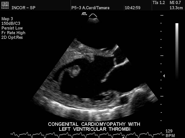
| This gray scale echo of the heart shows the left ventricle, anterior and posterior leaflets of the mitral valve, the aortic valve and the base of the aorta. Rounded echogenic fococus in the LV is reminiscent of thrombus. Courtesy Philips Medical Systems 33132 code cardiac heart echo LV MV aorta echogenic nodule thrombus congenital cardiomyopathy [dromano] |
Congenital Cardiomyopathy Dilated Left Ventricle and Thrombus in the LV |
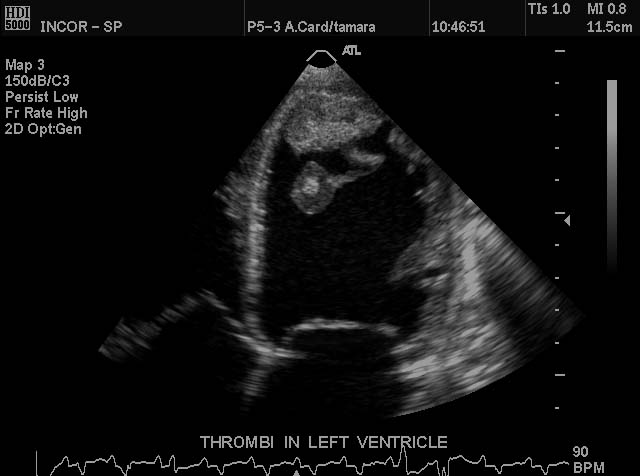
| This gray scale echo of the heart shows the left ventricle, and mitral valve. A rounded echogenic focus in the LV apex is reminiscent of thrombus. Courtesy Philips Medical Systems 33136 code cardiac heart echo LV apex mass echogenic thrombus cardiomyopathy imaging cardiac echo |
|
Structural Distrotion of the Pailary Muscles Complicated by Mitral Regurgitation |
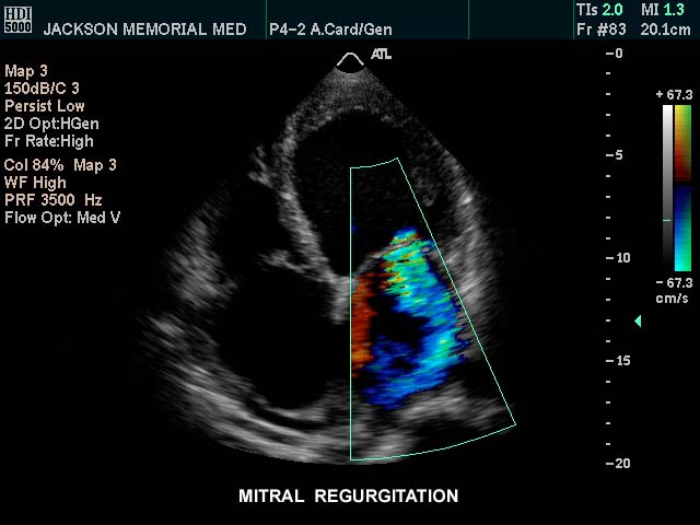
| This gray scale echo with color flow doppler of the heart shows a 4 chamber view. The regurgitant and turbulent jet of color through the mitral valve in to the left atrium is characteristic of mitral regurgitation in this patient with a cardiomyopathy. Courtesy Philips Medical Systems 33137 code cardiac heart echo MV MR cardiomyopathy see image 33131 [dromano] |
|
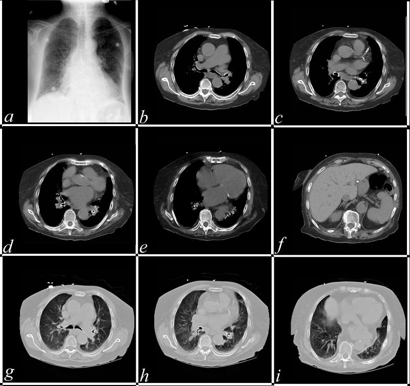
Ischemic Cardiomyopathy Atrial Fibrillation and Amiodorone Toxicity |
| This combination of CT images in this patient shows a plain film (a) with interstitial edema, followed by a series of images showing a large left PA, (b), calcific atherosclerotic coronary arterial disease (c), and 4 chamber cardiac enlargement (c,d,e) with apical thinning (e). The liver appears dense and measures 75 HU (f). Images g,h, and i reflect signs of congestion and interstitial edema. The likely diagnosis is a an ischemic cardiomyopathy, with congestive cardiac failure. What has caused the liver to be so dense? How do you link the abnormality to the cardiac failure?
Courtesy Scott Tsai 37106c 37106c |
|
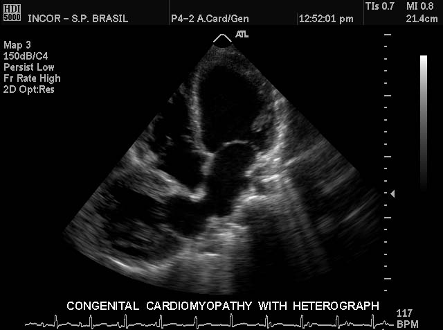
33131 |
| This gray scale echo of the heart shows 4 chambers of the heart of a pediatric patient witha congenital cardiomyopathy. To the right of the native heart is a heterograft piggy backed to enhance cardiac function. Courtesy Philips Medical Systems 33131 code cardiac heart echo congenital cardiomyopathy imaging cardiac echo |