
The Common Vein Copyright 2019
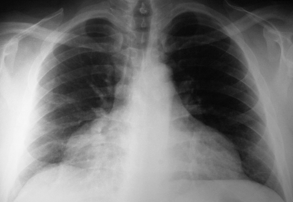
X-ray shows the typical appearance of a “water bottle heart” characteristic of a pericardial of a large pericardial effusion.
Ashley Davidoff MD
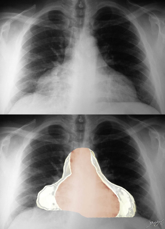
X-ray shows the typical appearance of a “water bottle heart” characteristic of a pericardial of a large pericardial effusion.
Ashley Davidoff MD
Atlas
| Effusion Type | T1W FSE | T2W FSE |
| Transudate | low | high |
| exudate | medium | high |
| proteinaceous | high | very high |
| acute hemorrhage | homogeneous high | homogeneous low |
| subacute or chronic hemorrhage | heterogeneous | subacute bright or dark
chronic usually dark |
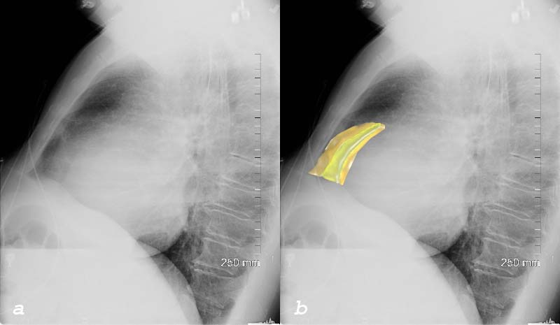
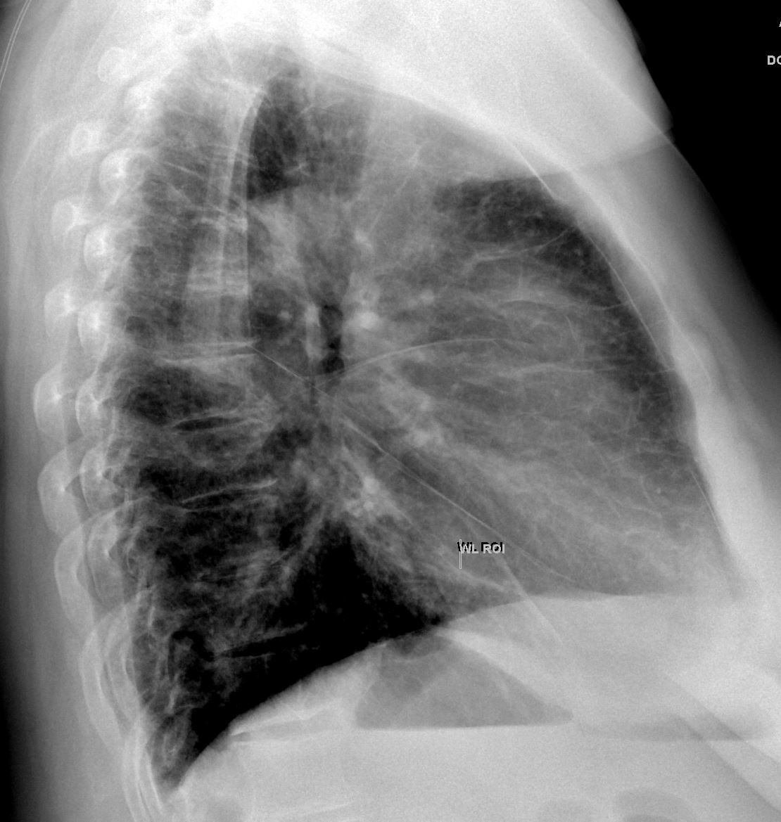
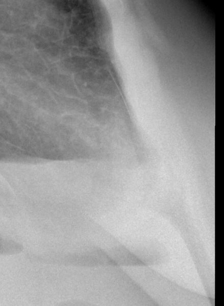
| Sandwich Sign – Oreo Cookie Sign of a Pericardial Effusion on Lateral CXR |
| 76031c01c heart cardiac pericardium pericardial effusion enlarged fluid between two fat layers sandwich sign visceral pericardium parietal pericardium epicardial fat pericardial fat plain film CXR plain X-ray Courtesy Rebecca Schwartz MD |
| Sandwich Sign of a Pericardial Effusion on Lateral CXR |
| 76031c01d heart cardiac pericardium pericardial effusion enlarged fluid between two fat layers sandwich sign visceral pericardium parietal pericardium epicardial fat pericardial fat plain film CXR plain X-ray Courtesy Rebecca Schwartz MD |
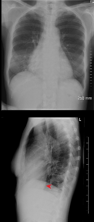
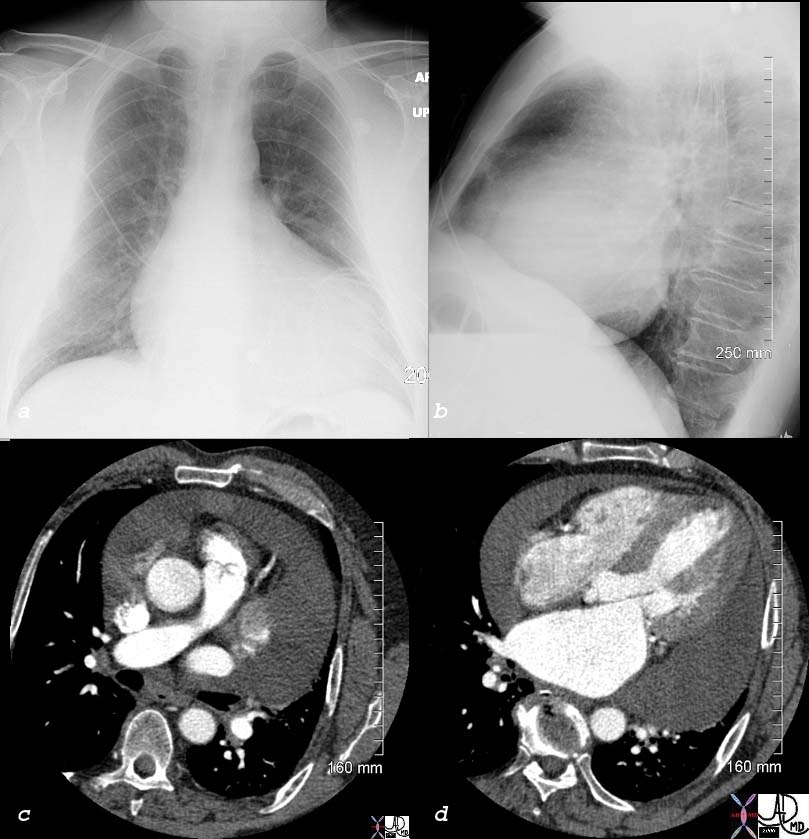
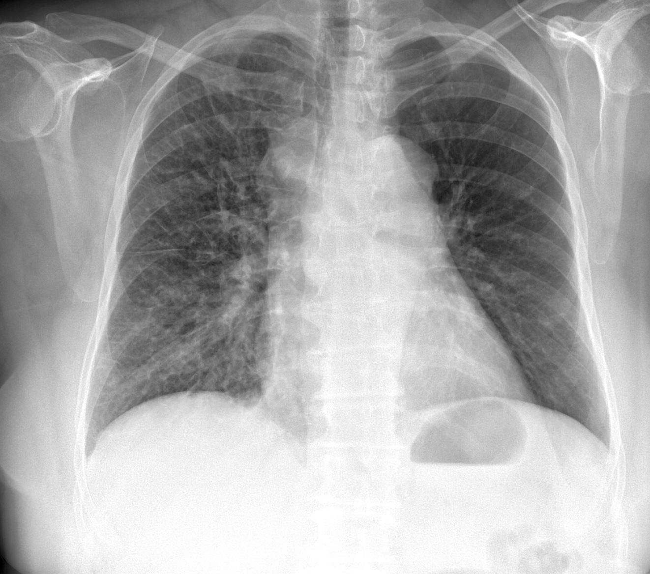
2 views of the chest of a 54 year old female with SLE and Sjogren’s syndrome show a slightly enlarged heart in the PA projection and an unusually prominent and rotund IVC (red arrowhead)
Ashley Davidoff MD
key words
pericarditis, pericardial effusion, SLE
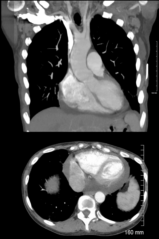
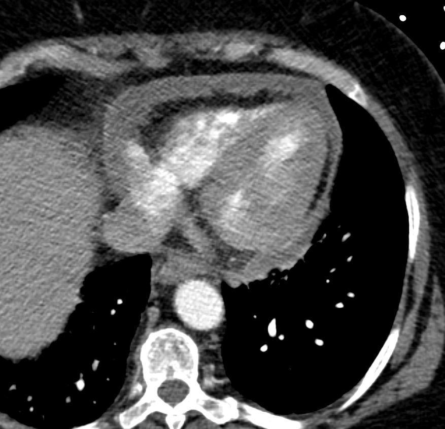
Coronal (above) and axial images CT showing the Rv and LV of a 54 year old female with SLE and Sjogren’s syndrome. A small pericardial effusion is noted accounting for the cardiomegaly on CXR.
Ashley Davidoff MD
key words SLE, pericarditis, pericardial effusion
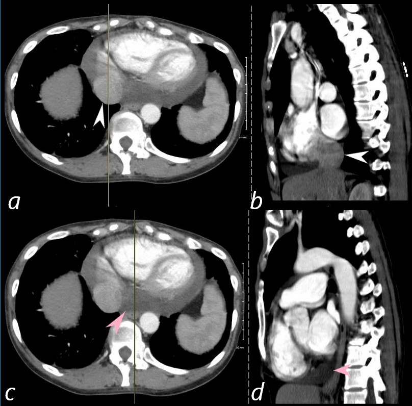
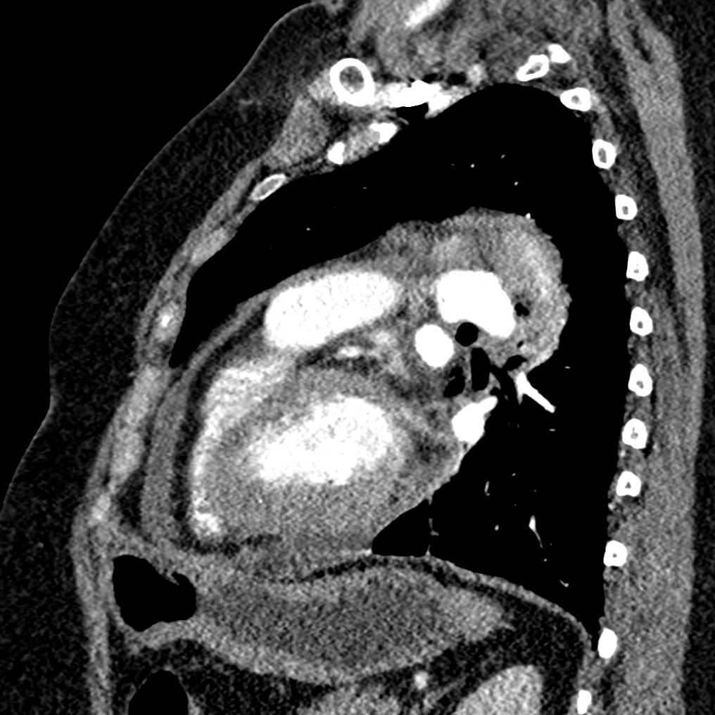
The axial image (a) and correlative sagittal image (b) show a prominent IVC (white arrowhead) accounting in part for the rotund and prominent IVC shadow on the CXR. Image c and d show that the pericardial effusion itself also contributes to the posterior shadow (pink arrowheads) The patient is a 54 year old female with SLE and Sjogren’s syndrome with active SLE and pericarditis and pericardial effusion.
Ashley Davidoff MD
key words SLE, pericarditis, pericardial effusion
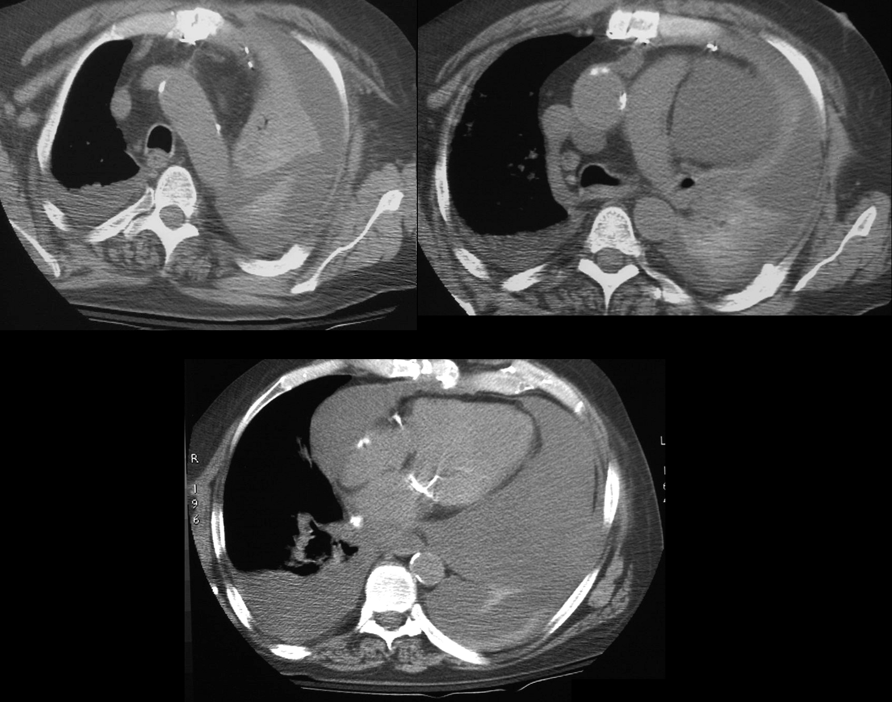
60 year old male s/p AVR presents with acute respiratory distress. CT scan shows total collapse of left lung, associated with a large effusion The atelectasis is either due to the associated effusion or an obstructing mucus plu. There is a large pericardial effusion with tamponade physiology by echo
Ashley Davidoff MD TheCommonVein.net
Oreo Cookie Sign
Ashley Davidoff MD TheCommonVein.net
Ashley Davidoff MD TheCommonVein.net
Ashley Davidoff MD TheCommonVein.net
Ashley Davidoff MD TheCommonVein.net
Ashley Davidoff MD TheCommonVein.net
Constriction
In CP, the size of the heart is usually normal; flattening of the right ventricle and curving of the interventricular septum to the left . The azygos vein and superior vena cava (SVC) are commonly dilated.
Web References
ACC – CXR and pericardial Disease