
82-year-old female with end stage renal failure, diabetes mellitus, CAD, s/p CABG x2 and AVR, severe mitral annular calcification, extending to the septum, s/p pacemaker, with associated mild mitral stenosis and significant mitral regurgitation. Severe aortic stenosis warranted AVR.
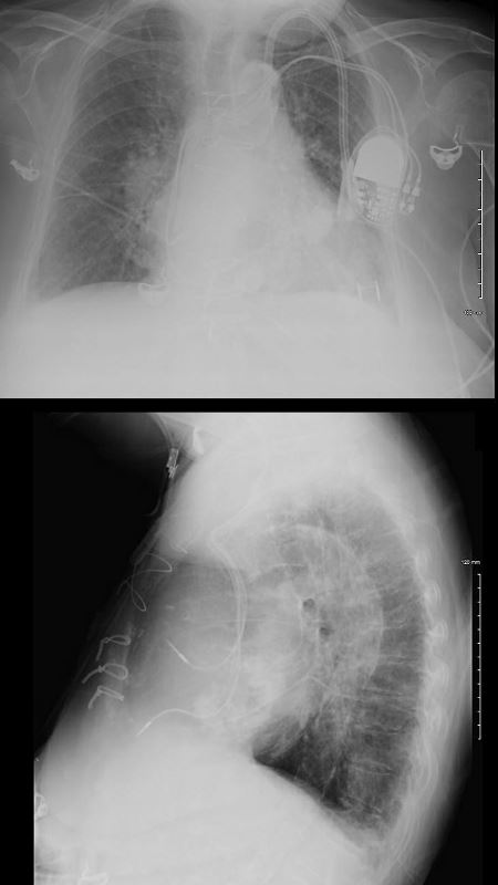
CXR shows severe mitral annular calcification, LVE, LAE and CHF.
Ashley Davidoff MD
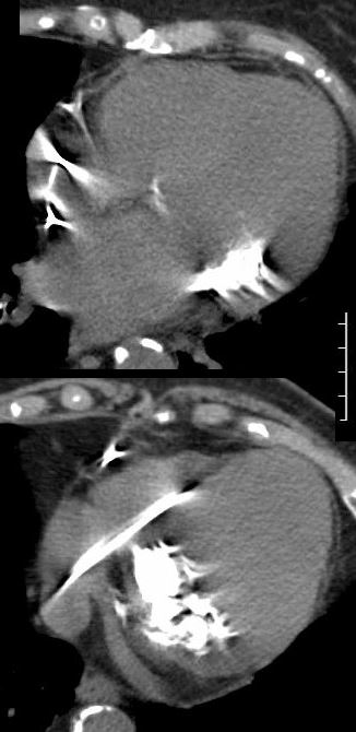
Axial CT through the mitral annulus shows severe mitral annular calcification extending into the ventricular cavity and the ventricular septum.
Ashley Davidoff MD
Sagittal reconstruction of the CT of the chest shows severe mitral annular calcification extending into the ventricular cavity and region of the septum. Note sparing of the anterior leaflet of the mitral valve.
Ashley Davidoff MD
Coronal reconstruction of the CT of the chest shows severe mitral annular calcification extending into the ventricular cavity and the ventricular septum.
Ashley Davidoff MD
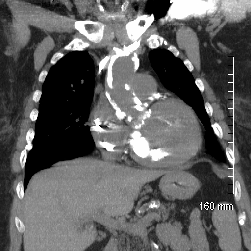
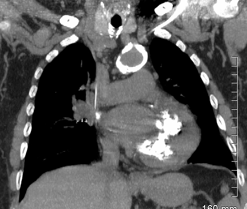
Coronal reconstruction of the CT of the chest shows severe mitral annular calcification extending into the ventricular cavity associated with aortic annular calcification.
Ashley Davidoff MD
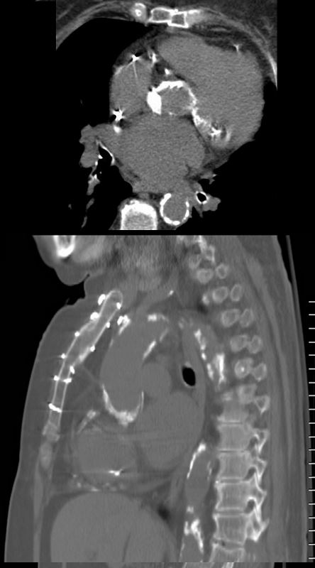
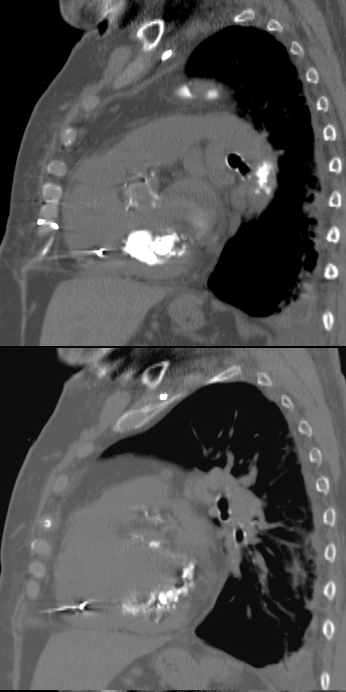
Axial image (above) and sagittal reconstruction of the CT (below) show aortic annular calcification.
Ashley Davidoff MD
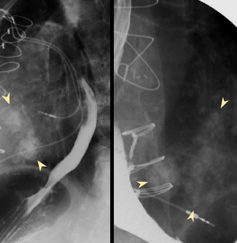
Barium swallow in LAO projection (left) and RAO (right) show heavy mitral annular calcification (yellow arrowheads).
Ashley Davidoff MD
