
Ashley Davidoff MD

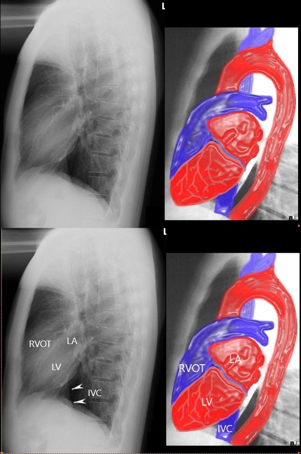
the anterior border of the chest is divided into thirds; 1/3 for the RVOT and 2/3 for the retrosternal air space
the posterior border of the heart is divided into thirds; 1/3 for the LA and 2/3 forthe LV.
the diaphragmatic border is divided into thirds; 1/3 for the LV and 2/3 for the rest of the diaphragm
Ashley Davidoff MD
15416C02Wlateral.8 rule of thirds
Left Ventricular Enlargement
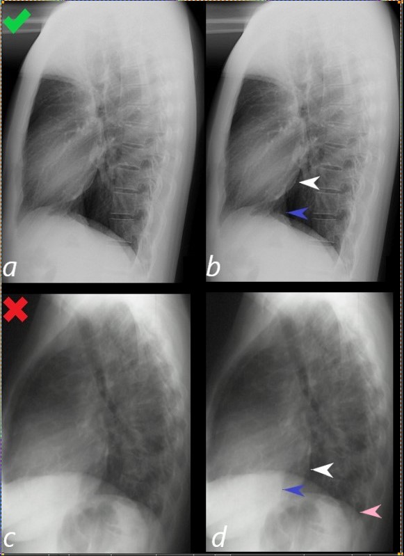
Lateral examination of a chest x-ray (CXR) shows the normal in the upper row (a,b) and the abnormal and enlarged in the bottom row (c,d).
The objective evaluation is based on the relative positioning and size of the LV (white arrowhead) in relation to the IVC, (blue arrowhead), and the left hemidiaphragm (pink arrowhead)
Ashley Davidoff MD
15416C02Wlateral LV01L.8
Right Ventricular Enlargement
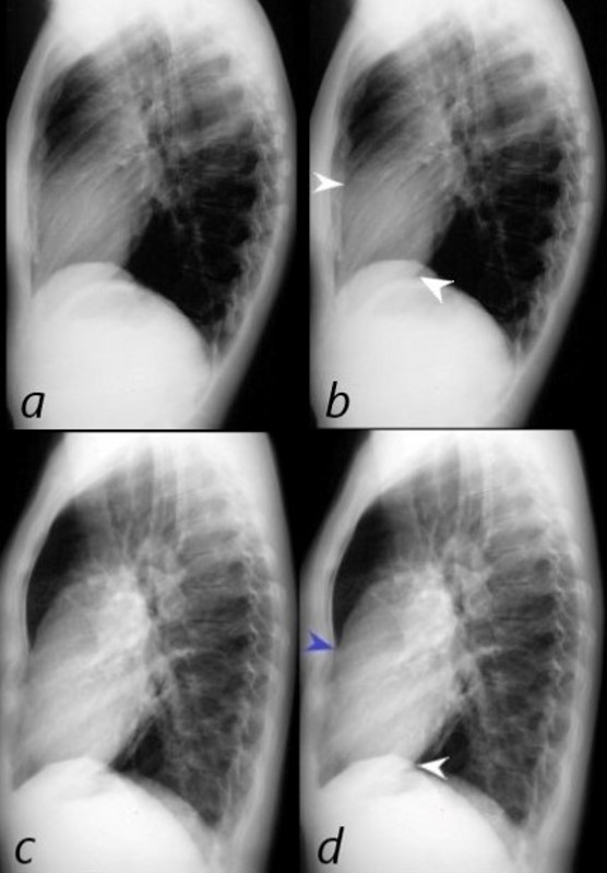
The normal lateral CXR (a,b), shows anterior and superior border of the heart (anterior white arrowhead) occupying 1/3 of the border between the sternomanubrial junction and the diaphragm.
The posterior and inferior white arrowhead shows the posterior border of the heart occupied by the RV taking up 1/3 of the distance of the diaphragm.
Images c and d represent left ventricular enlargement showing that the LV occupies about half the length of the diaphragm, (red arrowhead) while the retrosternal distance is unchanged and normal (white arrowhead).
Ashley Davidoff MD
Comparison between Left and Right ventricular Enlargement

The normal lateral CXR (a,b), shows anterior and superior border of the heart (anterior white arrowhead) occupying 1/3 of the border between the sternomanubrial junction and the diaphragm.
The posterior and inferior white arrowhead shows the posterior border of the heart occupied by the LV, taking up 1/3 of the distance of the diaphragm.
Images c and d represent left ventricular enlargement showing that the LV occupies about half the length of the diaphragm, (red arrowhead) while the retrosternal distance is unchanged and normal (white arrowhead).
Images e and f represent enlarged RV with the RV now occupying half the distance between the sternomanubrial junction and the diaphragm (blue arrowhead) while the distance occupied by the LV is still about 1/3 (white arrowhead)
Ashley Davidoff MD
 LATERAL EXAMINATION RVE AND LAE – MITRAL STENOSIS PULMONARY HYPERTENSION AND COR BOVINUM
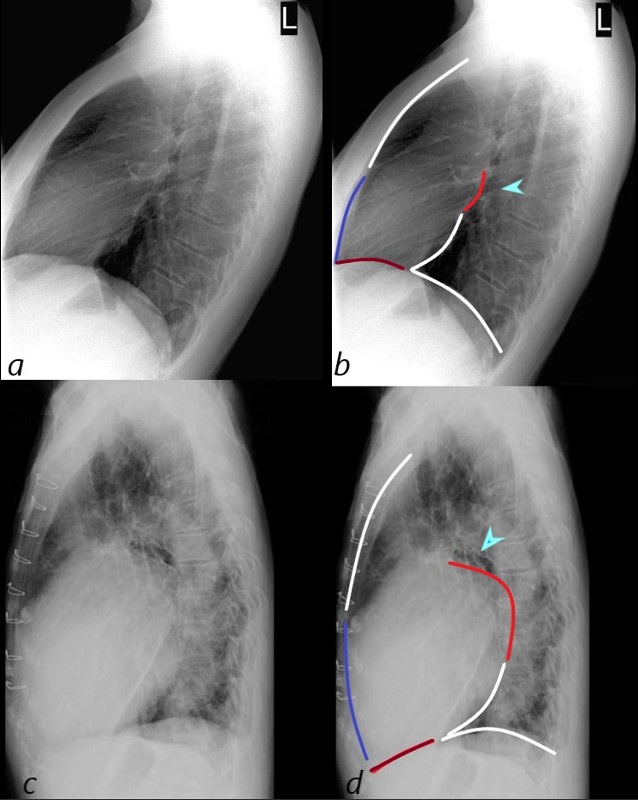
71 year old Asian female with rheumatic heart disease dominated by calcific mitral stenosis mild MR, moderate tricuspid regurgitation and secondary pulmonary hypertension.
The lateral examination (a, and b) show normal ratios of the length of the retrosternal air space, (upper anterior white line 2/3, and normal 1/3 length of the anterior RV border (blue line).
In images c and d the anterior overlay shows an increase in the ratio of the RV (navy blue-line to the upper 2/3 of the retrosternal space (white line) indicating RVE. Posteriorly, there is an increase ratio of the distance between the upper left atrial length (red – of posterior border of the left atrium (LA) and the lower border of the LV (white line) indicating LAE. In addition the straight left main bronchus (b – teal arrow) is shifted upward and is almost horizontal (d teal arrowhead) indicating LAE. Inferiorly the ratio of the length of the LV (maroon line) when compared to the rest of the diaphragm (white) is normal , indicating no evidence of LVE
Ashley Davidoff MD

Isolated Left Atrial Enlargement
This plain chest X-ray shows the classical features of an enlarged left atrium characterized by a double density on the right side of the heart (1), splaying of the carina (2), elevation and narrowing of the left main stem bronchus (3), prominent left atrial appendage (4), posterior displacement of the left main stem bronchus (5) as seen as the lateral examination and posterior bulging of the left atrium (6). These findings are consistent with mitral stenosis likely of rheumatic origin
Courtesy Ashley Davidoff MD copyright 2009 all rights reserved 15410c02.8s
IVC RA Junction

References and Links