Left Ventricle Hypertrophy with Hypertension (LVH)
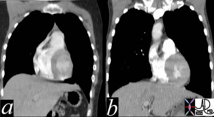
Coronal reformats through the chests of two different patients. They are approximately at the same level of the RVOT. The left image (a) is normal, while image b shows concentric thickening of the LV. Courtesy Ashley Davidoff MD 39088c01 code cardiac heart normal LVH LV hypertrophyThis series of axial and coronal CT scans (a,b,c,d,e) and coronal T1 weighted MRI scans show symmetric thickening of the left ventricle in this patient with LV hypertrophy. The MRI preceded the placement of the pacemaker noted in d and e. Courtesy Ashley Davidoff MD 39221c code cardiac heart LV myocardium thick concentric hypertrophy imaging radiology CTscanThis series of coronal CT scans (a,b,) T1 weighted MRI scans of the abdomen and lower chest show symmetric thickening of the left ventricle consistent with concentric LV hypertrophy. Courtesy Ashley Davidoff MD 39221c01.8 tags cardiac heart LV myocardium thick concentric hypertrophy imaging radiology CTscan