
Ashley Davidoff MD
130639L
75 year old female with findings consistent with cross fused ectopia. The left kidney is in intimate and inseparable contact with the right kidney and both lie in the right upper quadrantThey are both malrorated . The rightkidney is more superior and the pelvis points toward the right. The left kidney is lower and the pelvis points medially. There are 2 ureters. The right ureter enters the trigone on the right and the left on the left. 2 years later there is bilateral hydronephros as hydroureter of unknown etiology. Some mild reflux is noted to the right kidney on the cystogram
Ashley Davidoff MD
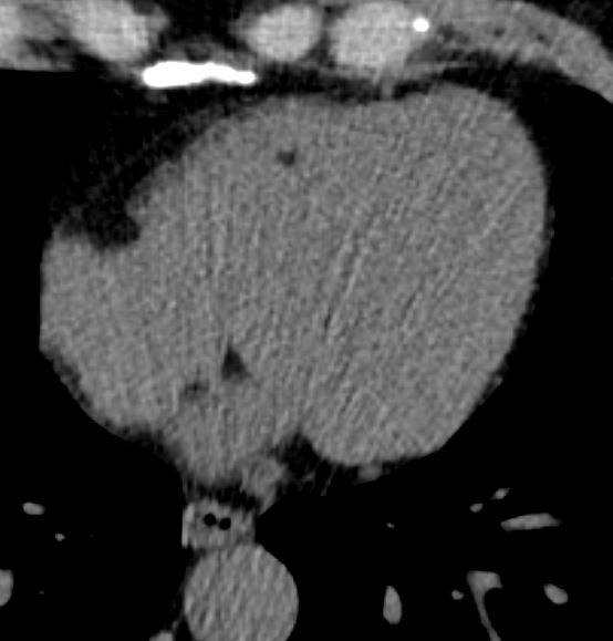
FAT IN THE MODERATOR BAND
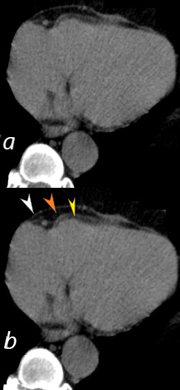
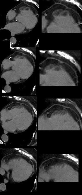
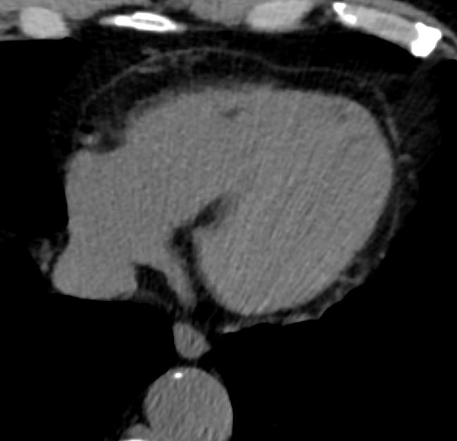
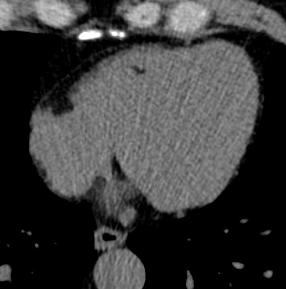
55 year old male with no known heart disease, with noted fat in the moderator band
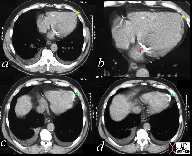
56 year old male with history of coronary artery disease. Axial CT through the heart shows apical curvilinear fat (yellow arrowheads, ( a and b) associated with apical myocardial dystrophic calcification (green arrowheads c and d) both indicating prior apical MI. In addition there mitral annular calcification (red arrowhead, b) and multifocal fatty deposits in the RV (white arrowheads, a and b) usually depicting age related degenerative changes,
Ashley Davidoff MD