
ASD Secundum
The Common Vein Copyright 2008
Ganesh Athappan MD Ashley Davidof f MD
Definition
ASD secundum is a congenital anomaly of fetal development characterized by a defect in the septum primum of atrial septum. It is the most common form of atrial septal defect.
The cause is a failure of closure of the ostium secundum and the result is a persistent communication between the right and left circulations at atrial level.
Structurally they are usually large single defects, but they may also be small and multiple.
Physiologically there is a shunting of blood from the left atrium to the right atrium .
Clinically they present in childhood with decreased exercise tolerance but they only present in 40-50 year olds with evidence of pulmonary hypertension. A systolic ejection murmur caused by increased flow across the pulmonary valve is heard over the second left intercostal space. Echocardiography confirms the diagnosis of a primum ASD. The direction of atrial level shunting can be detected by color Doppler.
Management is via transcatheter device closure, which is is the standard of care for secundum atrial septal defects .
The axial image through the heart at atrial septal level shows a flow void through a secundum ASD. The pink blood of the LA is shown shunting across and mixing with the blue blood of the enlarged RA.
01649b03 heart cardiac atrial septum secundum ASD secundum atrial septal defect secundum defect of the septum primum turbulence enlarged right atrium RAE right atrial enlargement congenital MRI Courtesy Ashley Davidoff MD copyright 2008
| ASD Secundum Total Absence of Septum Primum |
| The diagram shows a normal septum primum (white) and one with total deficiency of the septum primum (red) representing a large ASD secundum defect surrounded by the intact septum secundum.
01656b04.8s 01656b05j.8 Davidoff art Courtesy Ashley Davidoff MD copyright 2008 |
| Septum Primum |
| The septum primum is a membrane like structure surrounded by a rim of muscle seen in the first image, and diagrammed in the seconIt appears a s a fine membrane like structure and the body of the septum primum is intact except for the foramen ovale.
heart cardiac atrial septum septum primum septum secundum sinus venosus right atrium normal anatomy coronary sinus thebesian valve Eustachian valve coronary sinus triangle of Koch atrioventricular node A-V node grosspathology Courtesy Ashley Davidoff MD copyright 2008 all rights reserved 01671c.81s |
| ASD Secundum Total Absence of Septum Primum |
| The pathological specimen shows absence of the septum primum and a large defect that allows mixing of oxygenated blood from the left atrium (red) and deoxygenated blood (blue) from the SVC and IVC.
01661a02 heart cardiac interatrial septum fossa ovalis foramen ovale superior limbic band inferior limbic band septum secundum septum primum patent foramen ovale ASD secundum complete absence of the septum primum Davidoff art Courtesy Ashley Davidoff MD copyright 2008 |
| ASD Secundum Defect in the Septum Primum |
| The anatomical specimen shows a delicate septum primum held up with a forceps and a hole is noted in the middle characteristic of an ASD secundum. Unlike the first specimen this defect is a single hole surrounded by a partially intact septum primum. The fosaa ovalis is a wide defect above the septum primum. Note the membrane like appearance of the septum primum in the neonate. This seems to add credence to the theory of van Praagh that the septum primum originates from a the left venous valve of the sinus venosus.
heart atrial septum ASD secundum septum primum pathology Davidoff art copyright 2008 all rights reserved 01685b01c |
| ASD Secundum Defect in the Septum Primum Fenestrations |
| The anatomical specimen shows a delicate septum primum held up with a forceps and a hole is noted in the middle characteristic of an ASD secundum with two defects. The fosaa ovalis is a wide defect in this case just to the right (patients left ) and superior.
heart atrial septum ASD secundum septum primum pathology Davidoff art copyright 2008 all rights reserved 01658c |
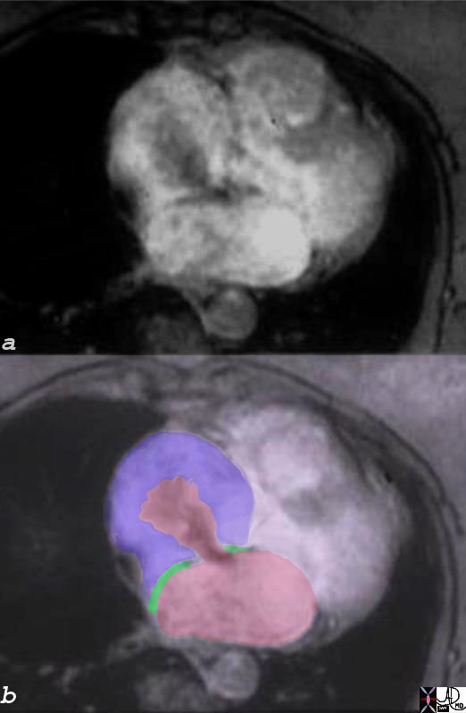
| MRI – ASD Secundum |
| The axial image through the heart at atrial septal level shows a flow void through a secundum ASD. The pink blood of the LA is shown shunting across and mixing with the blue blood of the enlarged RA.
01649b03 heart cardiac atrial septum secundum ASD secundum atrial septal defect secundum defect of the septum primum turbulence enlarged right atrium RAE right atrial enlargement congenital MRI Courtesy Ashley Davidoff MD copyright 2008 |
| Enlarged RA |
| The shunting of blood to the pulmonary circulation results in a volume overload on the RA and it becomes enlarged. The LA remains normal in size.
01639c01 right atrium heart enlarged RAE right atrial enlargement volume overload ASD secundum MRI T1 weighted Courtesy Ashley DAvidoff MD copyright 2008 |
| ASDsecundum and ASD primum |
| This anatomical specimen demonstrates both an secundum ASD surrounded by an intact septum secundum (green) and a primum ASD lying below the septum secundum overlying the tricuspid valve (orange).
00258c02 heart cardiac ASD secundum atrial septal defect of the primum type common atrioventricular cana’ common AVC defect superior limbic band inferior limbic band septum primum septum secundum grosspathology Courtesy Ashley Davidoff MD copyright 2008 |
| ASD secundum |
| Transesophageal echo using color flow doppler technology shows the heart at the atrial level with a jet of color going from left atrium (LA) in the near field to the right atrium (RA) in the far field. The defect is placed in the middle of the atrial septum making ASD II likely.
Courtesy Philips Medical Systems 33164 code heart LA RA atrial septum interatrial septal defect congenital imaging cardiac echo |
| Amplatz Device – ASD Repair |
| The PA and lateral CXR shows radioopaque Amplatz device in the atrial septum, better seen as a double disc inthe lateral exam. One component lies in the LA and the other in the RA and both rest and are secured toi the septum secundum.
48076 chest fx ASD rings interatrial rings dx atrial septal defect repaired with Amplatz device CXR plain film of chest X-ray Davidoff MD 48076 48071 |