
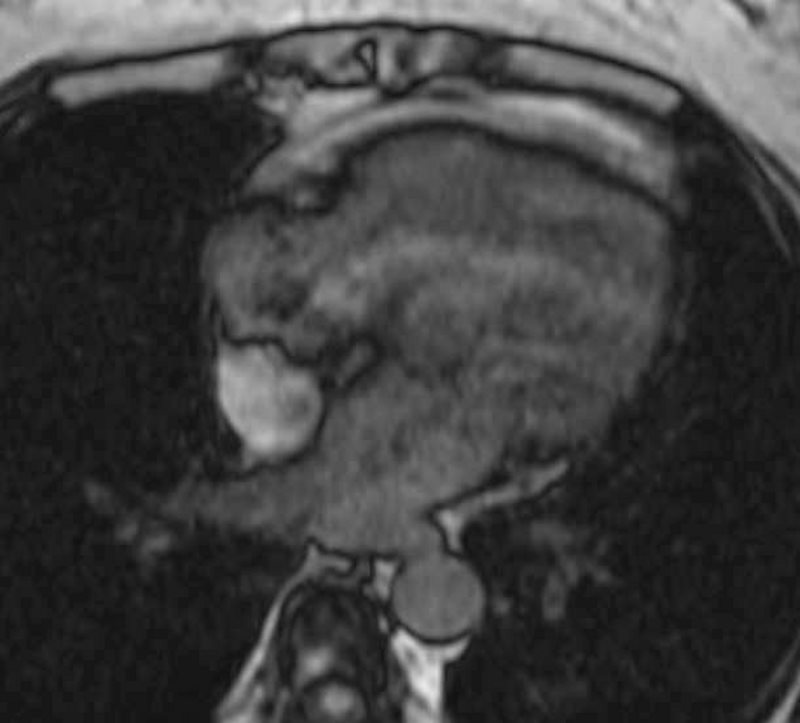
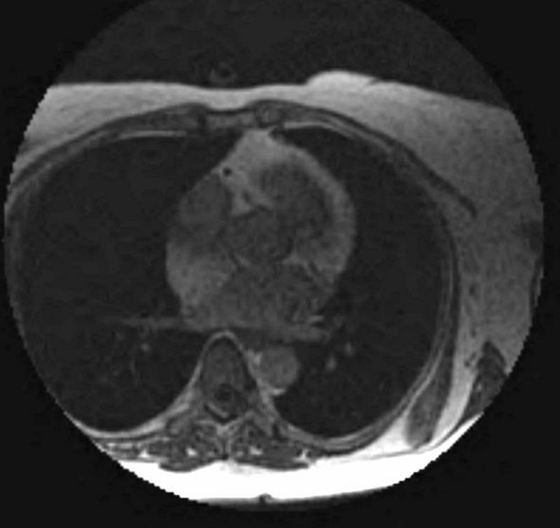
51-year-old female referred from echocardiography with diagnosis of a right atrial mass with suspected diagnosis of an atrial lipoma.
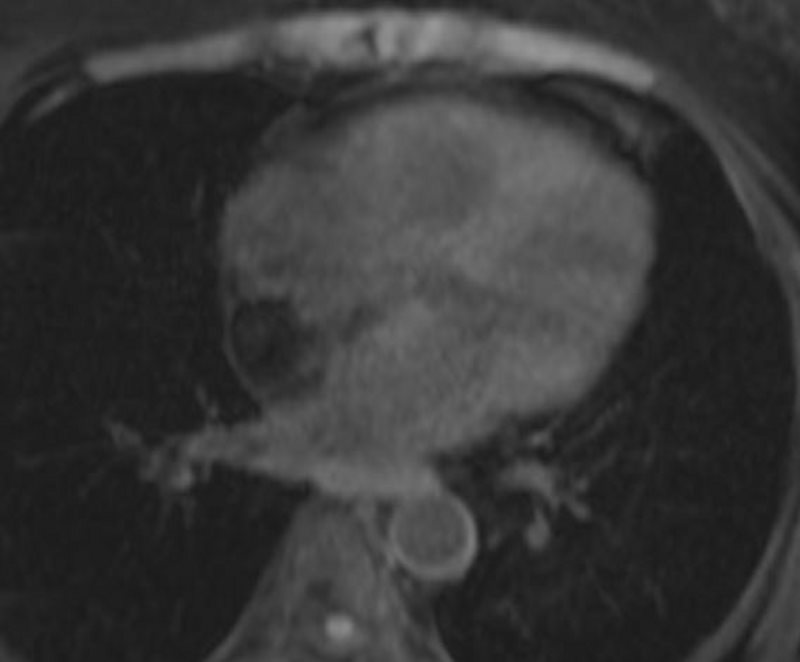
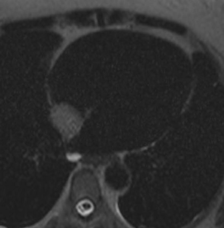
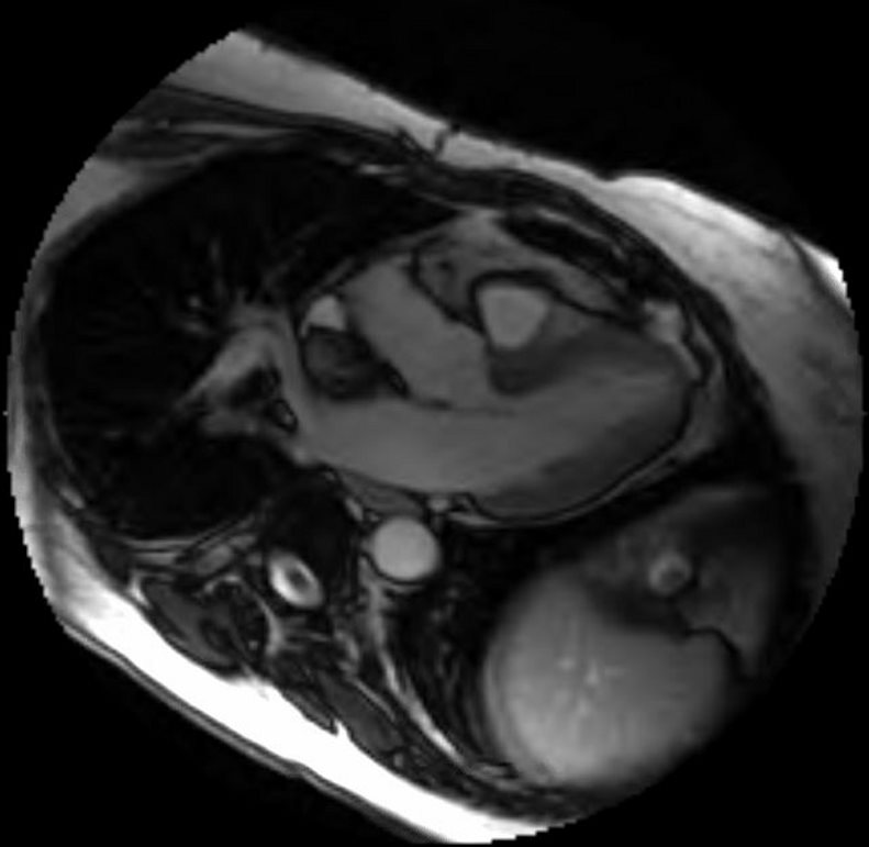
An MRCP confirms a lipomatous cardiac mass which is bright on T1 in phase, losing signal on the out of phase and fat sat sequences.
Ashley Davidoff MD
Ashley Davidoff MD
Ashley Davidoff MD
Ashley Davidoff MD
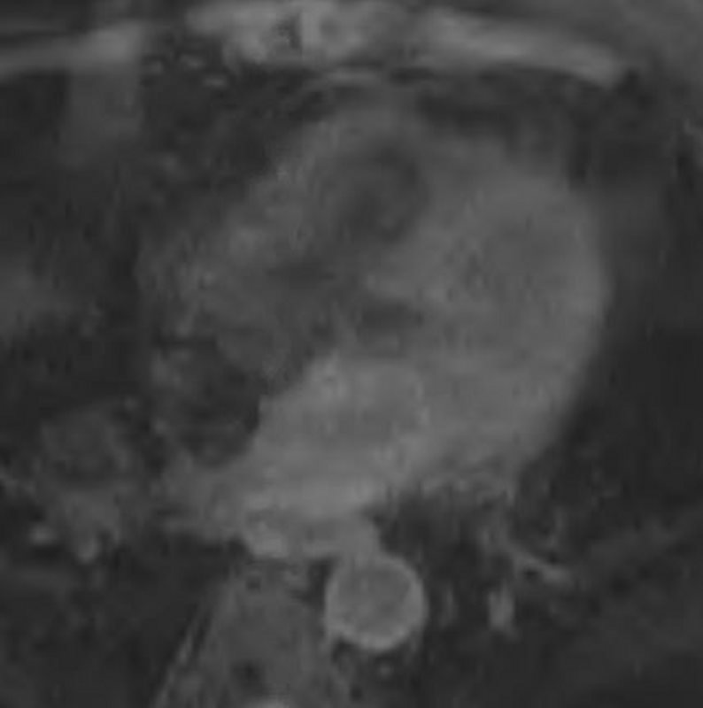
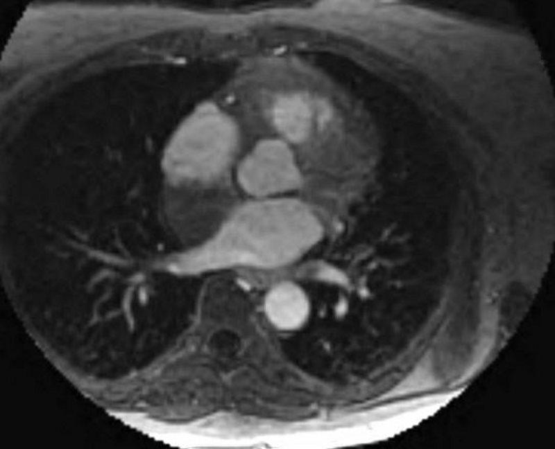
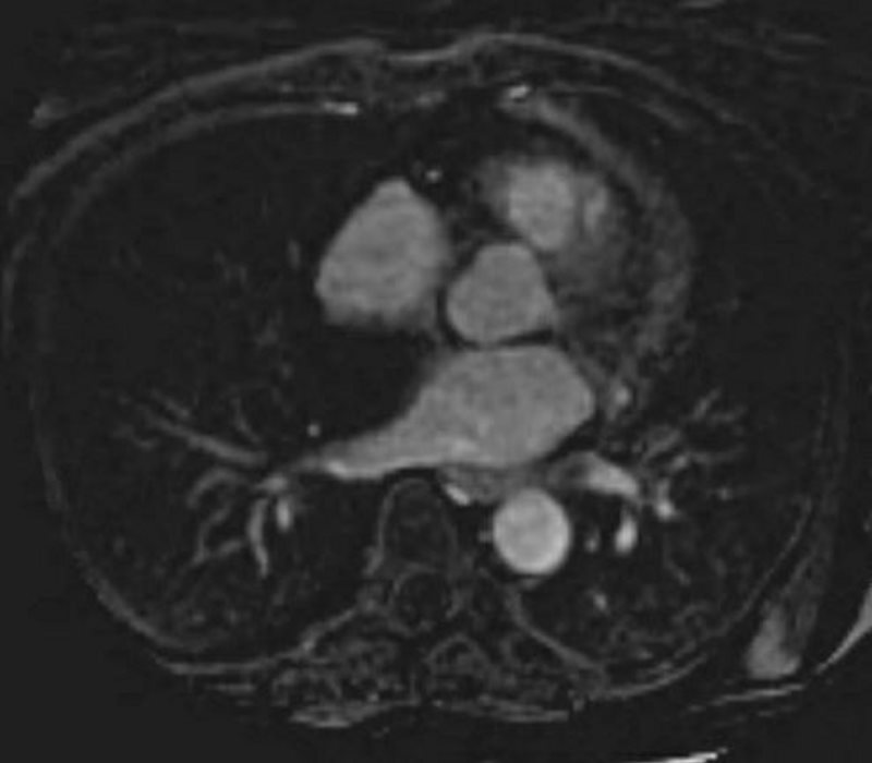
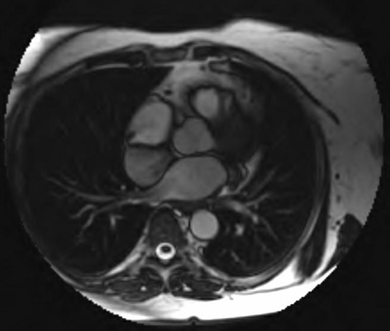
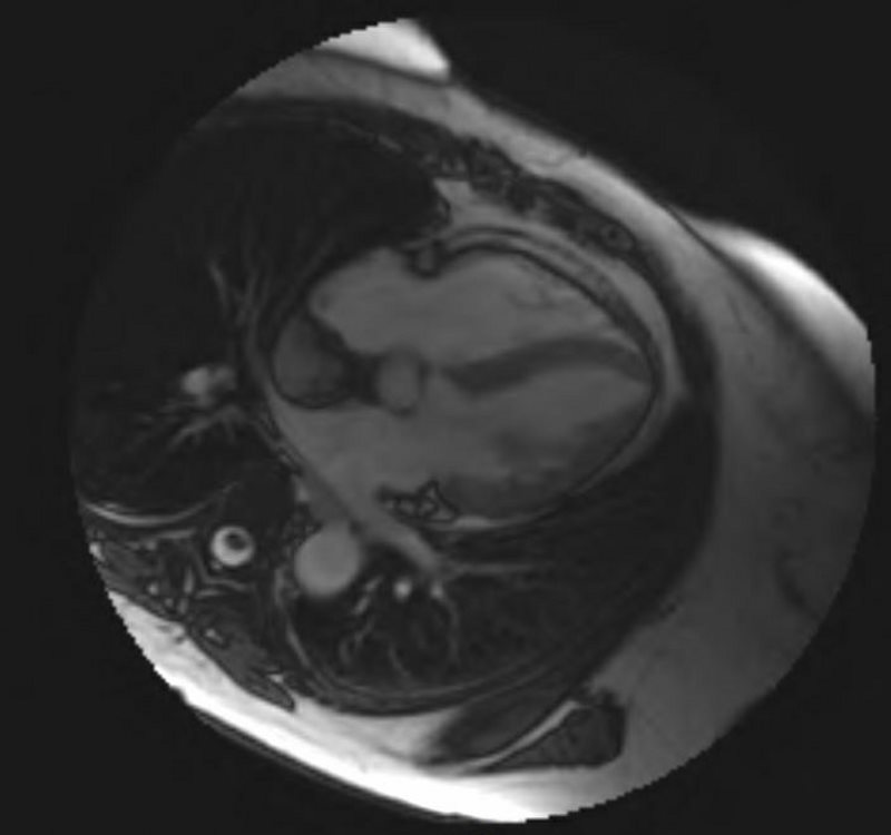
The dedicated cardiac MRI confirms the presence of a lipoma of the interatrial septum. There is no evidence of enhancement of the mass.
Ashley Davidoff MD
Ashley Davidoff MD
Ashley Davidoff MD
Ashley Davidoff MD
Ashley Davidoff MD
Ashley Davidoff MD
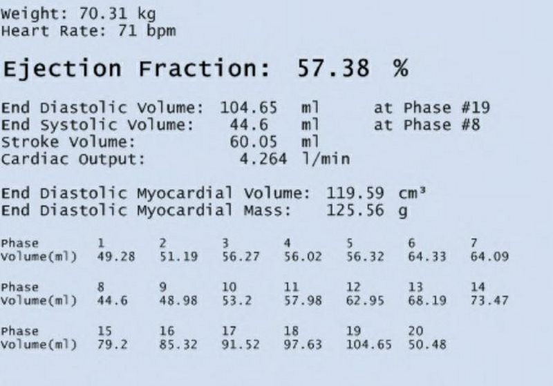
MRI – MEASUREMENTS
Ashley Davidoff MD
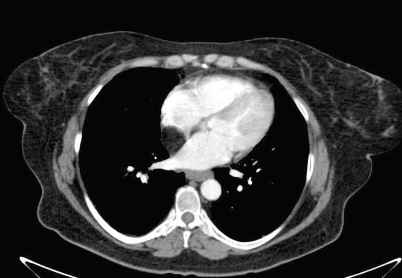
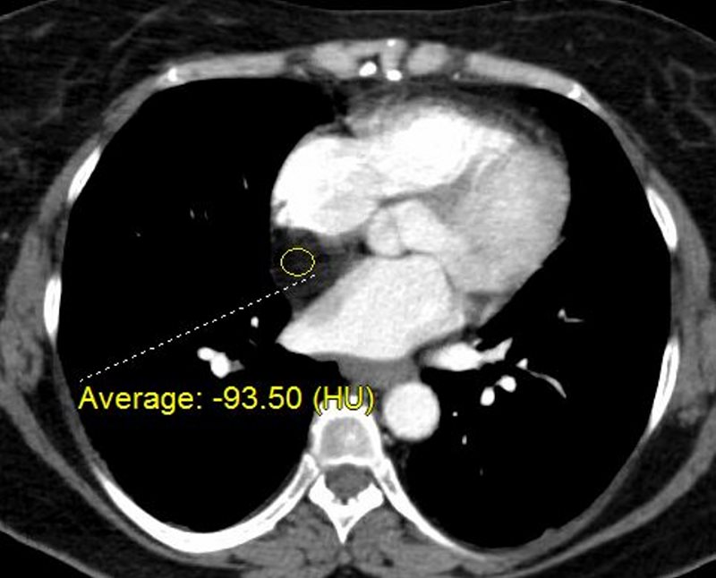
CT of the abdomen and pelvis, revealing the inferior aspect of the heart shows a lipomatous mass.
Ashley Davidoff MD
Ashley Davidoff MD
Ashley Davidoff MD
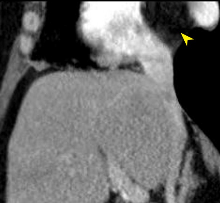
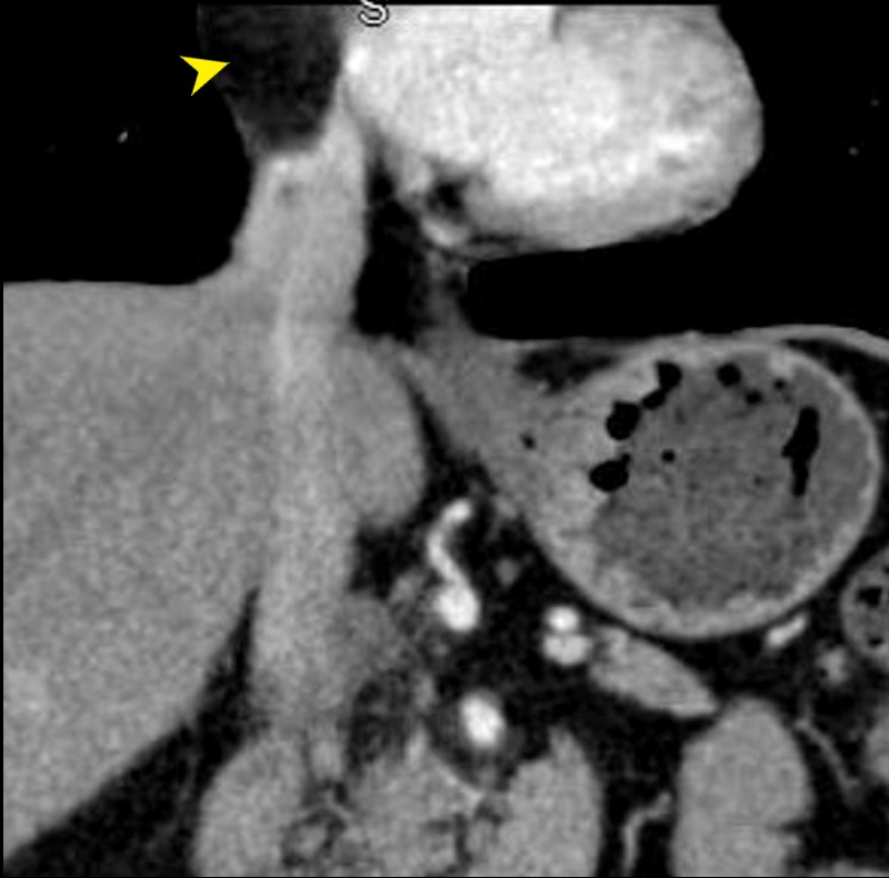
INTERATRIAL LIPOMA (yellow arrow)
Ashley Davidoff MD
These findings are all therefore consistent with lipoma of the interatrial septum