
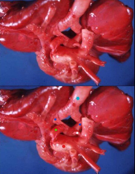
This is an autopsy specimen of a heart and lungs from a young patient with polysplenia and congenital heart disease who died following surgery. The important diagnostic feature in this specimen is the finding that both pulmonary arteries lie above the mainstem bronchi (dark blue dot)– ie bilateral hyparterial bronchi – a feature of bilateral left sidedness seen in polysplenia syndrome The image is taken from above showing the trachea (light blue dot) and the two-mainstem bronchi before the bronchi enter the lungs. Note the pink color of the lungs of this young patient, the surgical shunt from aorta to right pulmonary artery (green dot) , and the ductus from aorta to left pulmonary artery (white dot). The patient had a hypoplastic pulmonary valve with critical pulmonary stenosis.
Ashley Davidoff TheCommonVein.net 07236L
Links and References
-
Web
- TCV