
L – Transposition of the Great Vessels
Copyright 2009
Atlas
embryology bilateral conus DORV growth resorption normal mitral to aortic continuity transposition D transposition double outlet right ventricle
Ashley Davidoff
06394c02L01s
TheCommonVein.net
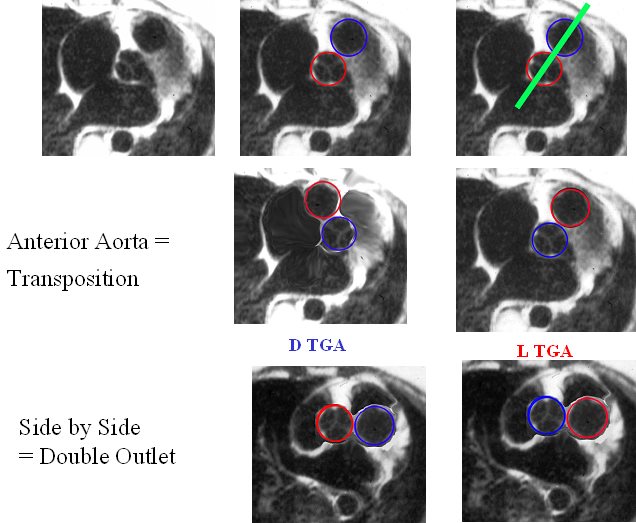
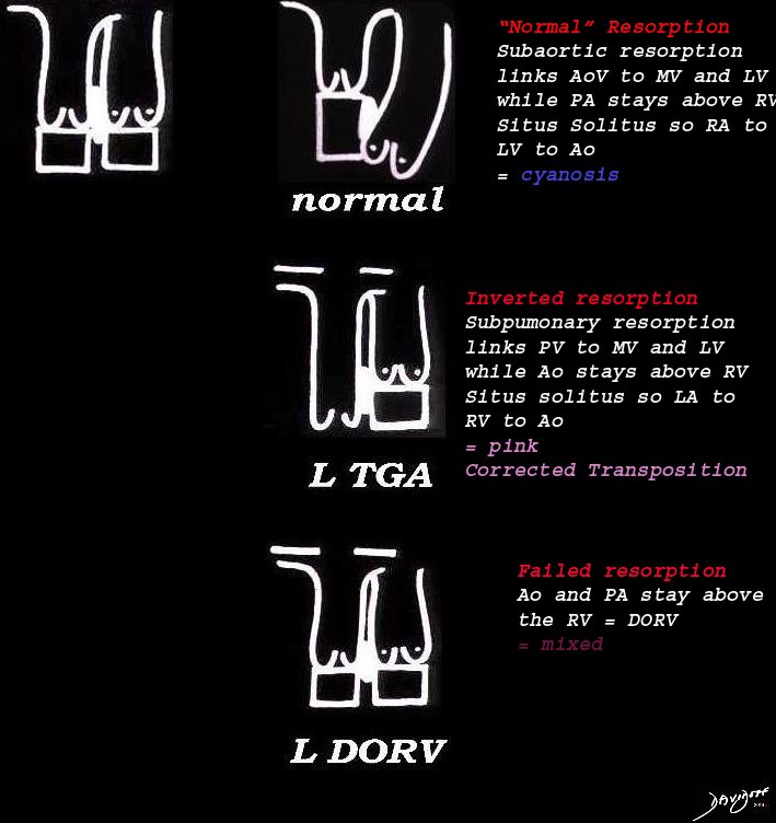
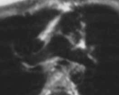
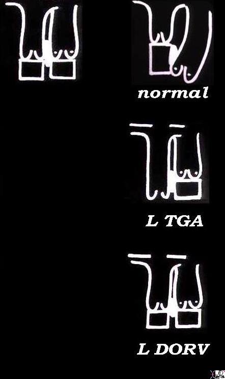
The image reflects the relationship of the aorta and pulmonary arteries in the normal patient, in DTGA, LTGA and DORV. In the normal patient with D loop the aorta (Ao) is posterior and to the right, and the pulmonary artery (PA) is anterior and to the right. In the patient with DTGA, the Ao is anterior and to the right and the PA is posterior and to the left. In an L loop the Ao is anterior and to the left and the PA is posterior and to the right. In double outlet right ventricle (DORV) the great vessels lie side by side and in DORV with a D loop the aorta is to the right and with an L loop the aorta is to the left
86778 01639 01639d02 01639d04 01639f03.jpg Anterior aorta = Transposition
Ashley Davidoff MD
TheCommonVein.net

eMediciene
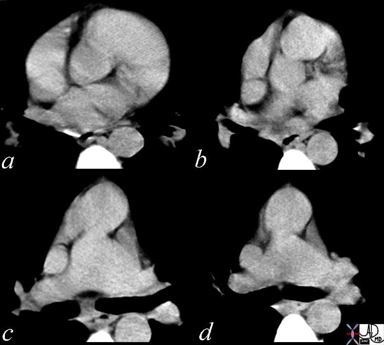
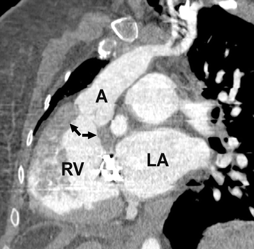
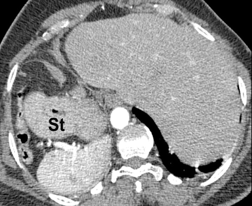
The axial CT reveals a findings characteristic of L transposition of the aorta with an anteriorly and leftward positioned aorta and posteriorly and rightward positioned pulmonary artery best seen in images c and d. In images a and b the distinction between aorta and pulmonary cannot be determined unless identifying which of the 2 vessels connects with the aortic arch
28999b01 heart cardiac aorta pulmonary artery RVOT conotruncal malformation LTGA L TGV transposition of the great vessels transposition of the great arteries corrected transposition position connection relation embryology CTscan Davidoff MD 28994 28995 28996 28997 28998 28999 MRI Ashley Davidoff MD TheCommonVein.net
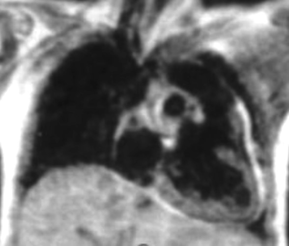
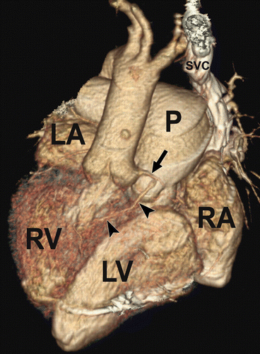
The left sided right ventrcle, receives oxygenated blood from the left sided left atrium, and gives rise to the left sided aorta. This configuration results in a soft tissue shape abnormality in the region of the AP window
TheCommonVein.net
The axial CT reveals a findings characteristic of L transposition of the aorta with an anteriorly and leftward positioned aorta and posteriorly and rightward positioned pulmonary artery.
07304b01 anterior aorta posterior pulmonary artery position connection subaortic conus L TGA L TGV L transposition of the great vessels L transposition of the great arteries levo leftward MRI Ashley Davidoff MD TheCommonVein.net
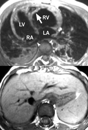
Fig. 4A —ECG-gated axial spin-echo T1-weighted MR images in patient with dextrocardia, situs solitus, and corrected transposition of great arteries (TGA). (Reprinted with permission from Reddy GP, Caputo GR. Diagnosis please: case 15. Radiology 1999; 211:709-710 [2]) Liver is on right and spleen is on left (bottom image), revealing situs solitus. Image through cardiac chambers (top image) shows discordant atrioventricular connections. Inferior pulmonary veins (arrowheads) drain to morphologic left atrium (LA). Left atrium is connected to morphologic right ventricle (RV), which is distinguished by presence of a moderator band (arrow). Morphologic right atrium (RA) is connected to morphologic left ventricle (LV). Ventricles are in L-loop configuration.
Read More: https://www.ajronline.org/doi/10.2214/AJR.06.1179
https://www.ajronline.org/doi/10.2214/AJR.06.1179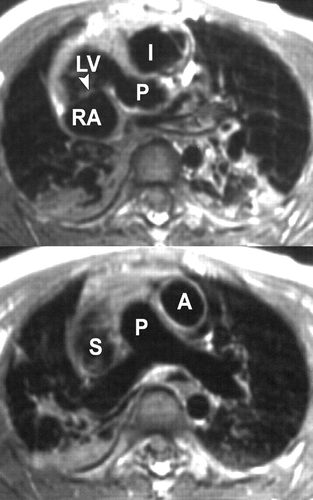
Read More: https://www.ajronline.org/doi/10.2214/AJR.06.1179
Read More: https://www.ajronline.org/doi/10.2214/AJR.06.1179
Read More: https://www.ajronline.org/doi/10.2214/AJR.06.1179
Read More: https://www.ajronline.org/doi/10.2214/AJR.06.1179
|
L – Loop DORV |
| 06394c02Ls embryology bilateral conus DORV growth resorbtion normal mitral to aortic continuity transposition L transposition corrected transposition double outlet right ventricle Davidoff art copyright 2009 all rights reserved |
References
- TCV
- Conotruncal Abnormalities
- Embryology
- Anatomy of the Right Ventricle
- Double Outlet Right Ventricle
- Double Outlet Left Ventricle
- Tetralogy of Fallot
- Transposition of the Great Vessels
- Truncus Arteriosus