
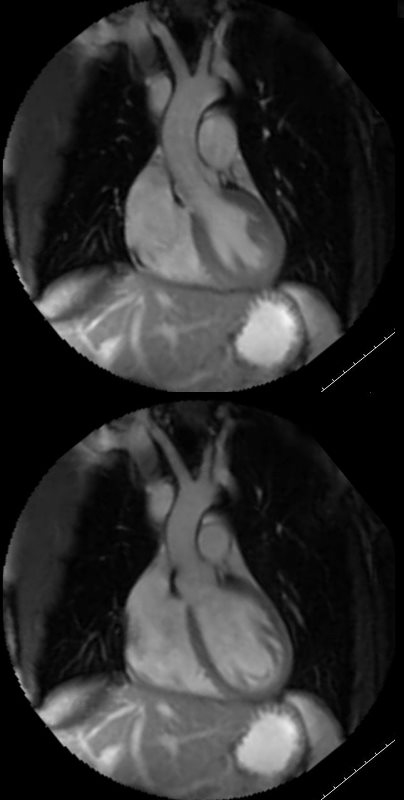
This is the MRI of a 19-year-old male who presented with syncope and the study was performed to identify a possible arrhythmogenic focus
White blood imaging of the LVOT view shows a normal sized ovoid LV in systole (above) and diastole (below). The walls appear normal thickness in the diastolic image, and the approximate residual volume of the LV at peak systole is about 1/3 the diastolic volume indicating an approximate ejection of 2/3 = 66% ejection fraction (EF).
Ashley Davidoff MD